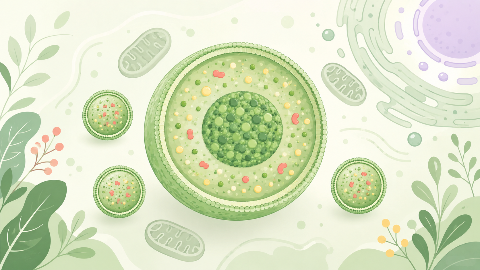
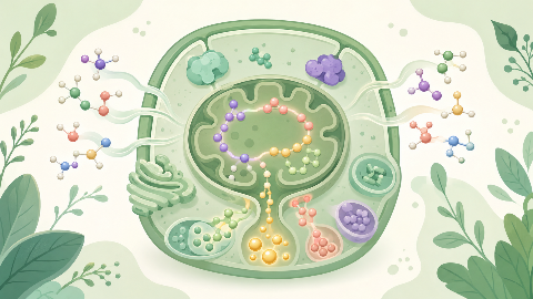
The peroxisomes
Discover peroxisomes, small cellular organelles that are key to the survival and adaptation of our cells! In this cell biology course, you'll explore their structure...
Osteology
The Osteology: The Occipital Bone course immerses you in the world of cranial bones. Discover their characteristics and location in the skull, as well as their importance for head movement. Experience an in-depth educational experience in the field of osteology!
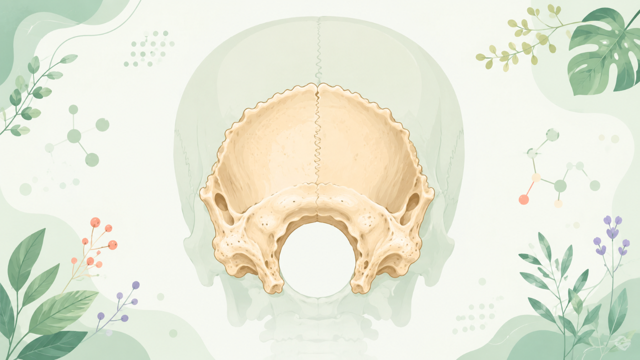
The occipital bone is the posterior and most caudal part of the cranial (skull) cavity, forming the base of the skull along with the sphenoid bone. This bone plays a significant role in providing attachment sites for various muscles and ligaments that stabilize and move the head. Understanding the anatomy and functional characteristics of the occipital bone is crucial in the field of osteology, as it contributes to the overall structural integrity and mobility of the skull.
The occipital bone develops from three distinct regions during embryonic development: the occipital part, the basilar part, and the exoccipital parts. These regions fuse together to form a single bone in adult life. The fusion process occurs approximately around the sixth month of gestation, although it may occasionally continue into the first few years after birth.
The occipital part arises from the rostral notochord and the mesoderm situated between the somites of the cervical region. It forms the major part of the adult occipital bone, including the basicranium and the greater and lesser wings of the sphenoid bone. The basicranium is a complex structure that houses the brainstem, cranial nerves, and the foramen magnum, which serves as the passageway for the spinal cord.
The basilar part develops from the ventral aspect of the mesoderm between the somites of the cervical region. In adult life, it contributes to the body and basioccipital portions of the occipital bone, as well as a significant portion of the sphenoid bone. The basioccipital is a prominent structure that articulates with the temporal bones laterally and forms the posterior part of the foramen magnum.
The exoccipital parts develop from the paraxial mesoderm located between the dorsal somites of the cervical region. These regions give rise to the paired supraoccipital and the paired occipital protuberances in adult life. The supraoccipital bones form the posterior part of the cranium, while the occipital protuberances contribute to the occipital ridge, which serves as an attachment site for the ligamentum nuchae and other muscles.
The occipital bone is divided into three primary regions: the basioccipital, the condyle, and the squama. Each region has specific characteristics and functions within the skull.
The basioccipital is a robust, quadrilateral structure that articulates with the temporal bones laterally and forms the posterior part of the foramen magnum. It bears two large, concave articular surfaces known as the condyles of the occipital bone, which facilitate movement between the skull and cervical vertebrae. The basioccipital also contains several openings (foramina) that transmit important vessels and nerves to various parts of the head.
The condyles are two large, round processes located at each extremity of the basioccipital. They articulate with the atlas (C1) and axis (C2) vertebrae, allowing for flexion, extension, and rotational movements of the head. The condylar region is covered in articular cartilage, which facilitates smooth movement between the skull and cervical vertebrae.
The squama is the largest and most dorsal part of the occipital bone. It consists of two parts: the greater wing and the lesser wing. The greater wing forms a prominent ridge known as the lambdoid suture, which articulates with the corresponding region of the parietal bones laterally. The lesser wing is smaller and thinner, forming the occipital ridge medially. Both wings contribute to the posterior part of the cranium, providing attachment sites for various muscles and ligaments.
The occipital bone plays a crucial role in maintaining the structural integrity of the skull and facilitating head movements. Injuries to this region can result in severe consequences, such as facial deformities, neurological dysfunction, or even death in extreme cases. Trauma to the occipital bone may occur during motor vehicle accidents, falls, or violent assaults.
In some pathological conditions, abnormal growths or deformations of the occipital bone can develop. These conditions include benign tumors (osteomas) and congenital malformations such as basilar invagination. Understanding the anatomy and potential complications associated with the occipital bone is essential for healthcare professionals working in neurosurgery, orthopedics, or trauma care.
The occipital bone is a critical component of the skull, playing a pivotal role in providing attachment sites for muscles, ligaments, and cranial nerves. Its development, structure, and regional anatomy are essential to understanding the overall function and integrity of the skull. The clinical relevance of the occipital bone underscores its importance in various medical specialties, particularly neurosurgery, orthopedics, and trauma care.
Do you think you know everything about this course? Don't fall into the traps, train with quizzes! eBiologie has hundreds of questions to help you master this subject.
Discover peroxisomes, small cellular organelles that are key to the survival and adaptation of our cells! In this cell biology course, you'll explore their structure...
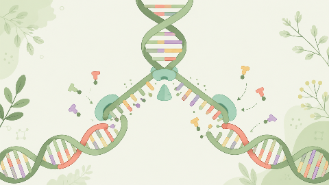
Discover how our DNA replicates with each cell division in this molecular biochemistry course: "DNA Replication." You'll learn the key steps in this crucial process...
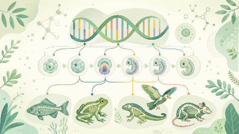
Learn about evolutionary developmental biology, the field that studies the mechanisms of embryonic development and their evolution at the molecular, cellular, and st...
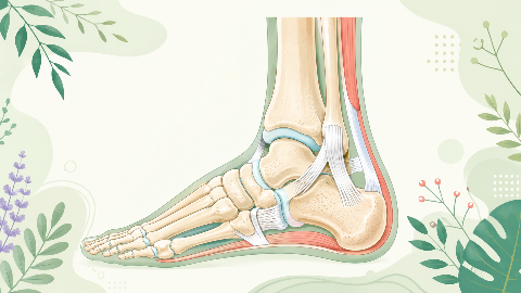
Discover the talonipedal joint in this syndesmology course: the bones, cartilage, and ligaments that enable the articulation of this complex joint. Learn to identify...
Discover amino acid metabolism in this exciting and technical course! You'll learn to identify the different types of amino acids and understand their essential role...
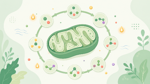
Discover the Krebs cycle: the central metabolic process that converts fatty acids into energy for the cell. Understand the steps in this process and learn how it int...
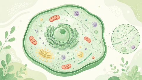
Dive into the intriguing world of Cellular Compartments! Explore the cytosol's structure, function, and dynamics, including its role in protein synthesis, energy pro...
