Osteology
course-show.h1-title
Discover the zygomaticus, a key facial bone for the structure of your face and the function of your lower jaw in this osteology course! Learn to identify and analyze its morphology and understand its role in the craniomandibular joint.
Introduction
The zygomatic bone, also known as the malar bone or cheekbone, is a complex and integral component of the skull in mammals. This bone forms part of the temporal bone and contributes significantly to the facial structure, particularly in humans where it is responsible for the prominence of the cheekbones. This comprehensive course delves into the various aspects associated with the zygomatic bone, providing a thorough understanding of its anatomical features, development, clinical significance, and evolutionary importance.
Anatomy of the Zygomatic Bone
Overview
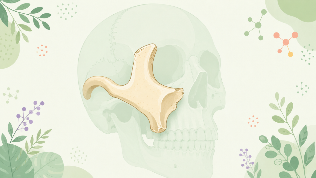
The zygomatic bone is one of the 14 bones that make up the cranium (skull). It is a flat, irregularly shaped bone located on either side of the face and forms part of the lateral wall of the orbit (eye socket) and the temporal region.
Structure
The zygomatic bone consists of three main parts:
- The square-shaped frontal process, which articulates with the frontal bone, forming the infraorbital margin.
- The malar process, which forms the prominent cheekbone and extends posteriorly to articulate with the zygomatic arch.
- The sphenoidal process, which articulates with the sphenoid bone and is located at the inferior and medial aspect of the zygoma.
Articulations
The zygomatic bone exhibits several important articulations:
- With the frontal bone, via the frontal process, forming the infraorbital margin and the root of the nasal bone.
- With the maxilla, via the zygomatico-maxillary suture, contributing to the floor of the orbit.
- With the temporal bone, via the zygomaticotemporal suture and the articular surface of the zygomatic process of the temporal bone, forming the zygomatic arch.
- With the sphenoid bone, via the sphenoidal process.
- With the palatine bone, via the palatine process of the maxilla (indirectly).
Development and Ossification
Embryology
The zygomatic bone originates from the first branchial arch during embryonic development. It develops from the mesenchyme and ossifies separately before fusing with other bones of the face during the latter stages of fetal life.
Ossification
Ossification of the zygomatic bone follows a centrifugal pattern, with two primary centers:
- The central center, which forms the body and becomes the frontal process in the fourth week of gestation.
- The peripheral center, which develops later at the margin of the future malar process. This center becomes the malar process by the sixth month, while the sphenoidal process ossifies from a separate center at the end of the first year.
Clinical Significance
Fractures and Trauma
Fractures to the zygomatic bone are common due to its location and structure. These injuries may lead to swelling, deformity, and impaired function. Depending on the severity and location of the fracture, treatment can range from conservative management with immobilization to surgical intervention for realignment or reconstruction.
Ophthalmic Concerns
Due to its proximity to the orbit, injuries to the zygomatic bone may cause orbital trauma, leading to complications such as enophthalmos (sunken eye), diplopia (double vision), and ptosis (drooping of the eyelid). In severe cases, surgical intervention may be necessary.
Evolutionary Significance
The zygomatic bone plays a crucial role in facial structure and expression, particularly in primates like humans. The enlargement and prominence of this bone have been linked to the development of complex social behaviors, communication, and species recognition among our ancestors. Furthermore, variations in zygomatic size and shape have been associated with differences in sexual selection, mating strategies, and overall reproductive success.
Conclusion
The zygomatic bone is a vital component of the skull, contributing significantly to facial structure, function, and evolutionary adaptations. Understanding its anatomy, development, clinical significance, and evolutionary importance provides a comprehensive foundation for students studying osteology and related fields.
Glossary
- Articulation: The region where two bones join together.
- Centrifugal pattern: A process in which the ossification begins at the center of the bone and moves outward toward the periphery.
- Cranium: The part of the skeleton that encloses the brain, consisting of 8 cranial and 14 facial bones.
- Embryology: The study of the development of an organism from its initial stages after fertilization to birth.
- Ossification: The process by which a bone hardens through the deposition of calcium salts in the matrix, forming bone tissue.
- Zygomatic arch: A curved bar-like structure formed by the articulation between the zygomatic bone and the temporal bones, providing support for the cheek and lower jaw.