Osteology
course-show.h1-title
Discover the coccyx, a small part of the human body essential for the stability and alignment of your spine. This course will deepen your knowledge of osteology by helping you identify its anatomical structure, understand its role in movement, and explore the abnormal variations that can occur. Broaden your understanding of the pelvic bones by taking this course on the osteology of the coccyx.
Introduction
The coccyx, also known as the tailbone or vertebra coccygea, is a unique and fascinating bone located at the terminal end of the vertebral column in humans. Although often overlooked in studies of human anatomy, this tiny yet vital structure plays an essential role in posture, balance, and pelvic stability. This course aims to provide an extensive and structured understanding of the coccyx within the field of osteology, encompassing its morphological, evolutionary, developmental, and clinical aspects.
Overview
In this course, we will embark on a journey to explore the coccyx's anatomy, evolutionary history, development during ontogeny, and clinical relevance in various contexts. The following chapters are organized hierarchically to facilitate a coherent and comprehensive learning experience:
Morphological Features of the Coccyx
- Anatomical Positioning
- Number of Segments
- Shape and Size
- Articulations and Ligamentous Connections
Evolutionary Perspective on the Coccyx
- Comparative Anatomy Across Vertebrates
- Evolutionary Transformations in Hominids
Developmental Aspects of the Coccyx
- Embryonic Origin and Formation
- Ontogeny of the Coccyx
Clinical Relevance of the Coccyx
- Injuries and Fractures
- Disorders and Diseases
- Clinical Examinations and Treatments
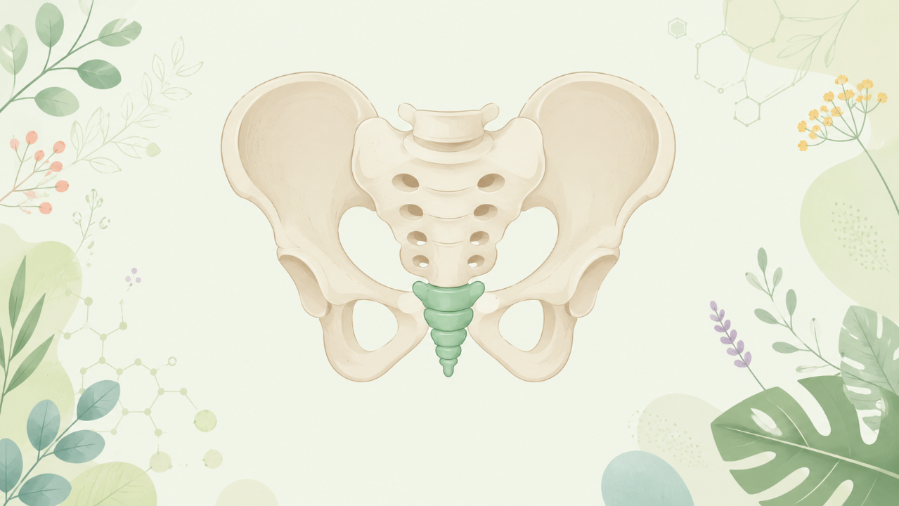
Morphological Features of the Coccyx
The coccyx is a triangular-shaped bone that consists of 3-5 fused vertebrae, with the number and size varying among individuals. The morphology of the coccyx can be further divided into several key features: anatomical positioning, number of segments, shape and size, and articulations and ligamentous connections.
Anatomical Positioning
The coccyx is located at the inferior part of the vertebral column, between the sacrum and the gluteus muscles. Its position allows it to play a crucial role in maintaining balance during sitting and standing postures while providing stability to the pelvis.
Number of Segments
Typically, the coccyx consists of 3-5 fused segments, with variations in the number being more common among females due to hormonal influences during pregnancy. The number of segments can affect the shape and size of the coccyx, as well as its overall functionality.
Shape and Size
The coccyx is triangular in shape, with its base directed posteriorly and apex pointing anteriorly. Its size varies among individuals, ranging from 2.5 to 4.5 cm in length, and it weighs approximately 30-50 grams.
Articulations and Ligamentous Connections
The coccyx articulates with the sacrum through the sacrococcygeal joint, which is a syndesmosis (synovial joint without articular cartilage). Additionally, the coccyx is connected to surrounding structures by various ligaments that help maintain its stability and mobility.
Evolutionary Perspective on the Coccyx
The evolution of the coccyx in vertebrates provides valuable insights into its role and function throughout history. This section will discuss comparative anatomy across vertebrates and examine the transformations that occurred during the evolution of hominids, leading to the formation of the human coccyx.
Comparative Anatomy Across Vertebrates
The tailbone is present in all vertebrates, serving various purposes such as locomotion, balance, and support for internal organs. In some species, the tail acts as an essential tool for grasping or propulsion. However, the number of segments in the tail varies among taxa, with humans possessing a small coccyx compared to other primates.
Evolutionary Transformations in Hominids
The reduction of the tailbone is a hallmark of hominid evolution, with the human coccyx being significantly smaller than that of our closest relatives, such as chimpanzees and gorillas. This adaptation likely occurred due to changes in locomotion patterns and posture during bipedalism, as well as for reasons related to reproductive advantages.
Developmental Aspects of the Coccyx
Understanding the developmental process of the coccyx provides essential insights into its origin, growth, and morphology. This section will examine embryonic origin and formation, as well as ontogeny of the coccyx during human development.
Embryonic Origin and Formation
The coccyx is derived from the caudal part of the notochord, which eventually gives rise to the vertebrae in this region. The number of segments in the tail varies due to processes such as segmentation and fusion during embryogenesis.
Ontogeny of the Coccyx
During fetal development, the coccygeal vertebrae differentiate from their respective somites and form a cartilaginous structure that eventually ossifies postnatally. The number of segments in the coccyx can increase during pregnancy due to hormonal influences but typically decreases after birth as segmentation and fusion occur.
Clinical Relevance of the Coccyx
The coccyx plays an essential role in maintaining balance, posture, and pelvic stability. However, it is also susceptible to injuries and disorders that can have significant impacts on a person's quality of life. This section will discuss common injuries, disorders, clinical examinations, and treatments associated with the coccyx.
Injuries and Fractures
Coccygeal injuries are often sustained due to traumatic events such as falls or motor vehicle accidents. Symptoms may include pain, swelling, and difficulty sitting. Treatment typically involves rest, pain management, and physical therapy in some cases.
Disorders and Diseases
Common disorders of the coccyx include coccydynia (pain in the tailbone) and coccygeal spondylolisthesis (dislocation of the coccyx). These conditions can be caused by various factors such as trauma, degenerative changes, or underlying medical conditions. Treatment depends on the severity of the condition but may include medication, physical therapy, or surgical intervention in severe cases.
Clinical Examinations and Treatments
Clinical examinations for coccyx-related issues involve palpation, range-of-motion tests, and imaging studies such as X-rays or MRIs. Treatment strategies are tailored to the specific condition and may include conservative measures like rest, pain management, and physical therapy, as well as more invasive approaches like surgery in severe cases.