Osteology
course-show.h1-title
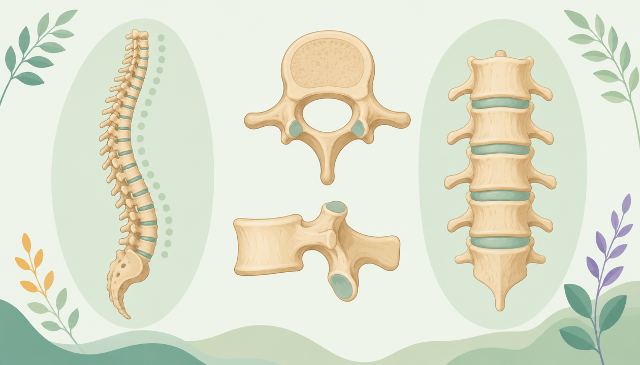
Discover the central skeletal system of vertebrates: the vertebrae. In this osteology course, we will explore the morphology and function of each segment of this bone chain, essential for locomotion, protection of the spinal canal, and postural maintenance. Discover how these varied structures enable a wide variety of forms and lifestyles in vertebrates.
Introduction
The vertebral column, or spinal column, is a complex and integral part of the skeletal system in the human body. This article aims to provide a comprehensive and detailed overview of the vertebrae, their classification, morphology, articulation, development, and clinical significance.
Vertebral Anatomy
Classification
The vertebral column consists of 33 individual vertebrae that can be classified into four groups based on their structure and function: cervical, thoracic, lumbar, and sacral/coccygeal vertebrae.
Cervical Vertebrae (C1-C7)
- Atlas (C1) : The first cervical vertebra, also known as the atlas, is a ring-like bone that supports the head and allows for rotation of the head during movement.
- Axis (C2) : The second cervical vertebra, or axis, bears a unique odontoid process which articulates with the atlas to enable flexion and extension movements of the neck.
Thoracic Vertebrae (T1-T12)
- Shape: Thoracic vertebrae are characterized by their box-like shape due to the presence of a large body, transverse processes, and strong, posterior, articular facets for articulation with the ribs.
Lumbar Vertebrae (L1-L5)
- Shape: Lumbar vertebrae have larger bodies and shorter neural spines compared to thoracic vertebrae, allowing for increased flexibility in the lower back.
- Number: Typically, the human body has five lumbar vertebrae.
Sacral (S1-S5) and Coccygeal Vertebrae (Co1-Co4)
- Sacrum: The sacrum is a triangular bone formed by the fusion of five vertebrae (S1-S5). Its anterior surface is concave, while its posterior surface is flat and convex.
- Coccyx (Tailbone): The coccyx is composed of four fused vertebrae (Co1-Co4) and has a flattened, triangular shape. It provides attachment sites for muscles and ligaments in the pelvic region.
Vertebral Morphology
General Characteristics
Each individual vertebra shares several common features:
- A body: The central part of a vertebra that forms the main mass.
- Pedicles: Bony projections arising from either side of the body, connecting to the lamina.
- Lamina: A bony plate formed by the fusion of the posterior halves of two vertebral neural arches, providing protection for the spinal cord.
- Spinous Process: A long, upright projection from the midline of the lamina, palpable through the skin in many regions of the back.
- Transverse Processes: Smaller bony projections extending laterally and slightly superiorly from each side of the vertebral body. They help to attach muscles, ligaments, and joints.
- Superior Articular Facets: Oval-shaped surfaces on the superior (upper) part of each vertebra, articulating with the inferior articular facets of the vertebra above it.
- Inferior Articular Facets: Similar to superior articular facets but located inferiorly, articulating with the superior articular facets of the vertebra below it.
Distinguishing Characteristics
Cervical Vertebrae
- Atlas (C1): The atlas has no body or spinous process due to its ring-like structure, but it has two laterally projecting transverse processes and superior and inferior articular facets on each half of the ring.
- Axis (C2): The axis has a large anterior arch with an odontoid process articulating with the posterior arch of the atlas. It also has transverse processes and superior and inferior articular facets like other cervical vertebrae.
Thoracic Vertebrae
- Shape: Thoracic vertebrae are wider than they are tall, with a tall neural spine and wide, flat bodies that articulate with the ribs.
- Vertebral Notches: These notches, present on either side of the body, provide sites for the heads of the ribs to attach.
Lumbar Vertebrae
- Shape: Lumbar vertebrae are taller than they are wide, with a shorter neural spine and larger bodies compared to thoracic vertebrae.
- Lamina Fusion: The lamina of the first lumbar vertebra (L1) is fused anteriorly, while the remaining lumbar vertebrae have separate laminae.
Sacral and Coccygeal Vertebrae
- Fusion: The sacrum and coccyx are formed by the fusion of multiple individual vertebrae, resulting in a single bone for each region.
- Shape: The sacrum is triangular, while the coccyx is cylindrical.
Vertebral Articulation
The articulation between adjacent vertebrae occurs at the inferior and superior articular facets of each vertebra. This allows for flexion, extension, lateral bending, and rotation of the spine. The intervertebral discs, located between individual vertebrae, contribute to this mobility by absorbing compression forces and allowing limited motion.
Vertebral Development
The vertebrae develop from embryonic mesodermal tissue called somites, which give rise to the sclerotome, a specific group of cells that differentiate into bones. The sclerotome condenses and gives rise to the ossification centers for each individual vertebra, eventually forming mature bone through endochondral ossification.
Clinical Significance
The vertebral column is susceptible to various injuries, degenerative changes, and pathologies such as herniated discs, osteoarthritis, and fractures. Understanding the anatomy, morphology, and function of each individual vertebra can aid in the accurate diagnosis and treatment of these conditions.
Conclusion
The vertebral column plays a crucial role in providing structural support, protecting the spinal cord, and allowing mobility in the upper body. Understanding its anatomy, morphology, development, and clinical significance is essential for healthcare professionals involved in diagnosing and treating related conditions.