Histology
course-show.h1-title
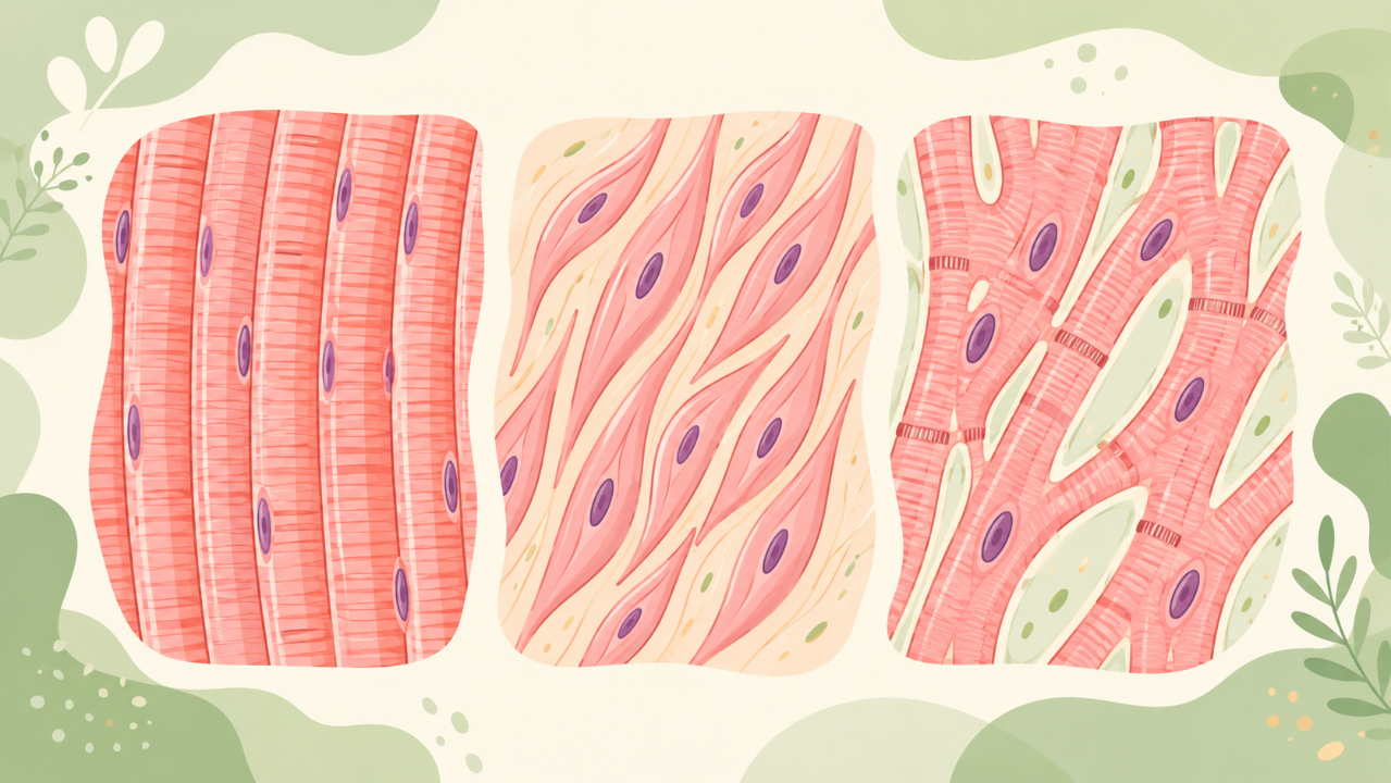
Discover the key differences between striated (skeletal and cardiac muscles) and smooth (smooth muscle) muscle tissue. Examine their ultrastructural structure, function, and role within the body.
Introduction
Muscle tissue is a specialized form of animal tissue that has the primary function of contracting to produce force and motion. In this course, we will delve into the comparative histology of three major types of muscle tissues: striated (skeletal and cardiac), smooth, and cardiac muscles. We will examine their structural and functional differences, as well as the cellular organization that allows them to carry out their respective roles in the body.
Striated Muscles
Skeletal Muscles
Anatomy
Skeletal muscles are attached to bones and are responsible for movement of the skeleton. They are composed of elongated, multinucleated muscle fibers that are connected to the skeleton via tendons at each end. The individual muscle fibers are themselves comprised of numerous myofibrils, which contain repeating units called sarcomeres.
Sarcomere Structure
A sarcomere is the functional and structural unit of a striated muscle fiber. It consists of thick filaments (myosin) arranged in parallel to thin filaments (actin). The thick and thin filaments are connected by a complex of proteins called the Z-disk, which forms the repeating pattern seen in skeletal muscles under microscopy.
Regulation of Contraction
The regulation of contraction in striated muscle fibers is highly coordinated by an intricate system of nerves and chemical signals. Action potentials travel along the nerve fibers to reach motor endplates, where they release acetylcholine, which binds to nicotinic receptors on the muscle fiber membrane. This causes an influx of calcium ions into the cytoplasm, triggering the contraction process through a series of molecular interactions.
Cardiac Muscles
Anatomy
Cardiac muscles are responsible for the rhythmic contractions of the heart that pump blood throughout the body. They differ from skeletal muscles in their continuous, involuntary contraction pattern. Unlike skeletal muscle fibers, cardiac muscle fibers are typically shorter and branched, allowing them to form a complex three-dimensional network within the heart wall.
Sarcomere Structure
The sarcomere structure of cardiac muscles is similar to that of skeletal muscles, with thick and thin filaments arranged in parallel. However, certain differences exist, such as the presence of additional proteins (e.g., titin) that contribute to the unique mechanical properties of cardiac muscle.
Regulation of Contraction
The regulation of contraction in cardiac muscles is also different from skeletal muscles. Instead of relying on acetylcholine, cardiac muscles are stimulated by an internal pacemaker, located within the sinoatrial node. This rhythmic electrical activity travels throughout the heart muscle via specialized conduction pathways, initiating a series of coordinated contractions that propagate throughout the heart.
Smooth Muscles
Anatomy
Smooth muscles are found in organs and tissues where constant or intermittent contraction is required for their proper function. Examples include blood vessels, stomach, intestines, and reproductive organs. Unlike striated muscles, smooth muscle fibers are spindle-shaped and uninucleate.
Sarcomere Structure
The sarcomeres of smooth muscles have a similar structure to that of striated muscles but with some key differences. For example, the Z-disk is less prominent in smooth muscle fibers, and there is more overlap between the thick and thin filaments. These structural variations contribute to the unique contractile properties exhibited by smooth muscles.
Regulation of Contraction
The regulation of contraction in smooth muscles differs from both striated and cardiac muscles. Smooth muscle cells are stimulated by a variety of chemical signals, including hormones (e.g., epinephrine) and neurotransmitters (e.g., acetylcholine). These signals act on intracellular receptors to initiate a chain of events that ultimately lead to the activation of myosin ATPase, which powers muscle contraction.
Conclusion
In conclusion, understanding the comparative histology of muscle tissue allows us to appreciate the remarkable diversity and specialized functions exhibited by these critical tissues in our bodies. From the coordinated movement of skeletal muscles to the rhythmic contractions of cardiac muscles and the continuous activity of smooth muscles, each type plays a vital role in maintaining the integrity and function of our organism as a whole.