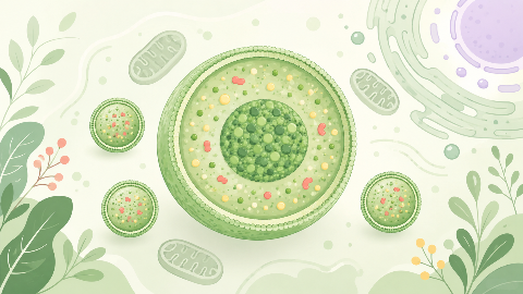
The peroxisomes
Discover peroxisomes, small cellular organelles that are key to the survival and adaptation of our cells! In this cell biology course, you'll explore their structure...
Histology
Discover the secrets of nervous tissue in this fascinating histology! You'll learn the structure and function of neurons, as well as the different types of nervous tissue. You'll also follow the evolution of this tissue throughout embryonic and adult development.

Histology, the study of the microscopic structure of tissues in multicellular organisms, provides an essential foundation for understanding the functions and interactions between various biological structures at different scales. One of the most critical and complex types of tissue within the body is the nervous tissue. This article aims to provide a comprehensive overview of the various aspects associated with nervous tissue, focusing on its structure, function, development, and pathology.
Nervous tissue is one of the four fundamental types of tissues in animals (the others being epithelial, connective, and muscle tissues). It consists primarily of specialized cells called neurons and supportive cells known as glia. Neurons are responsible for transmitting electrical and chemical signals throughout the body, enabling communication between different parts of the nervous system and other organs.
Neurons can be broadly classified into three main types based on their shape and function: sensory neurons, interneurons, and motor neurons. Sensory neurons receive information from various receptors in the body and transmit this information to the central nervous system (CNS). Interneurons are responsible for processing and integrating information within the CNS itself. Motor neurons transmit signals from the CNS to effector cells (such as muscles or glands), thereby enabling muscular contraction, secretion, and other responses.
The basic structure of a typical neuron consists of three main parts: the cell body, dendrites, and axons.
The cell body (soma) is the largest part of the neuron and contains the nucleus and other organelles essential for protein synthesis, metabolism, and growth. The cell membrane surrounding the cell body forms a boundary between the intracellular and extracellular environments.
Dendrites are specialized extensions of the neuron that receive incoming signals from other neurons or sensory receptors. They are highly branched and have multiple spines, which increase the surface area available for receiving inputs. The dendrites convey this information to the cell body, where it can be processed.
Axons are long, slender extensions of the neuron that transmit electrical signals away from the cell body to other neurons or effector cells. They have a myelin sheath, which insulates and speeds up the transmission of these signals. The axon terminates at a synapse, where it releases neurotransmitters to signal another cell.
Glial cells, or glia, provide support and protection for neurons within the nervous system. They outnumber neurons in the human brain by approximately tenfold. There are three main types of glial cells: astrocytes, oligodendrocytes, and microglia. Each plays a distinct role in maintaining the integrity and function of the nervous tissue.
Astrocytes are the most abundant type of glial cell in the brain. They surround neurons, providing structural support and protecting them from mechanical damage. Astrocytes also play a vital role in regulating neurotransmitter levels, maintaining the blood-brain barrier, and contributing to the formation of the myelin sheath around axons.
Oligodendrocytes are responsible for producing the myelin sheath that surrounds many axons in the central nervous system. This insulation facilitates faster and more efficient transmission of electrical signals along the axon.
Microglia are the resident immune cells of the brain, acting as scavengers and sentinels. They play a critical role in maintaining homeostasis within the nervous tissue by phagocytosing cellular debris, modulating inflammation, and participating in synaptic pruning during development and adult life.
The development of the nervous system begins during the early stages of embryonic development, with the formation of the neural plate and its subsequent folding to form the neural tube. This process is essential for the proper differentiation and organization of neurons and glial cells within the CNS. Abnormalities in this process can result in various congenital neurological disorders.
Several diseases and conditions specifically affect the nervous tissue, including Alzheimer's disease, Parkinson's disease, multiple sclerosis, and epilepsy. Each of these disorders results from a combination of genetic, environmental, and immunological factors that lead to dysfunction or degeneration of neurons and glial cells within the CNS.
Nervous tissue is a complex and fascinating structure that enables communication between various parts of the body, making it essential for coordinated responses and maintaining homeostasis. By understanding the basic structure, function, development, and pathology of nervous tissue, we can gain valuable insights into both normal physiological processes and disease states affecting this crucial organ system.
Do you think you know everything about this course? Don't fall into the traps, train with quizzes! eBiologie has hundreds of questions to help you master this subject.
Discover peroxisomes, small cellular organelles that are key to the survival and adaptation of our cells! In this cell biology course, you'll explore their structure...
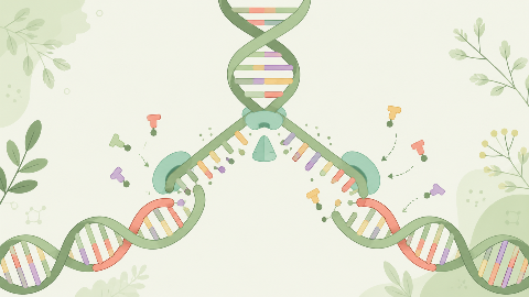
Discover how our DNA replicates with each cell division in this molecular biochemistry course: "DNA Replication." You'll learn the key steps in this crucial process...
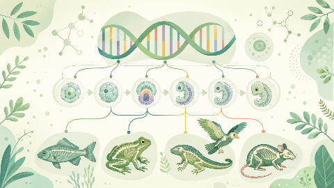
Learn about evolutionary developmental biology, the field that studies the mechanisms of embryonic development and their evolution at the molecular, cellular, and st...
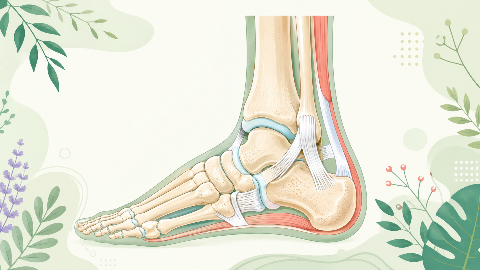
Discover the talonipedal joint in this syndesmology course: the bones, cartilage, and ligaments that enable the articulation of this complex joint. Learn to identify...
Discover amino acid metabolism in this exciting and technical course! You'll learn to identify the different types of amino acids and understand their essential role...

Discover the Krebs cycle: the central metabolic process that converts fatty acids into energy for the cell. Understand the steps in this process and learn how it int...

Dive into the intriguing world of Cellular Compartments! Explore the cytosol's structure, function, and dynamics, including its role in protein synthesis, energy pro...