Myology
course-show.h1-title
Discover the muscles of the head and neck: an overview of the structures that govern our chewing, speaking, and facial movements.
Introduction
The human head and neck region house a complex array of muscles responsible for various essential functions, including facial expressions, mastication, deglutition, respiration, speech, and maintaining posture. This comprehensive course provides an in-depth exploration of the muscles of the head and neck, offering a detailed understanding of their structure, function, innervation, and clinical significance.
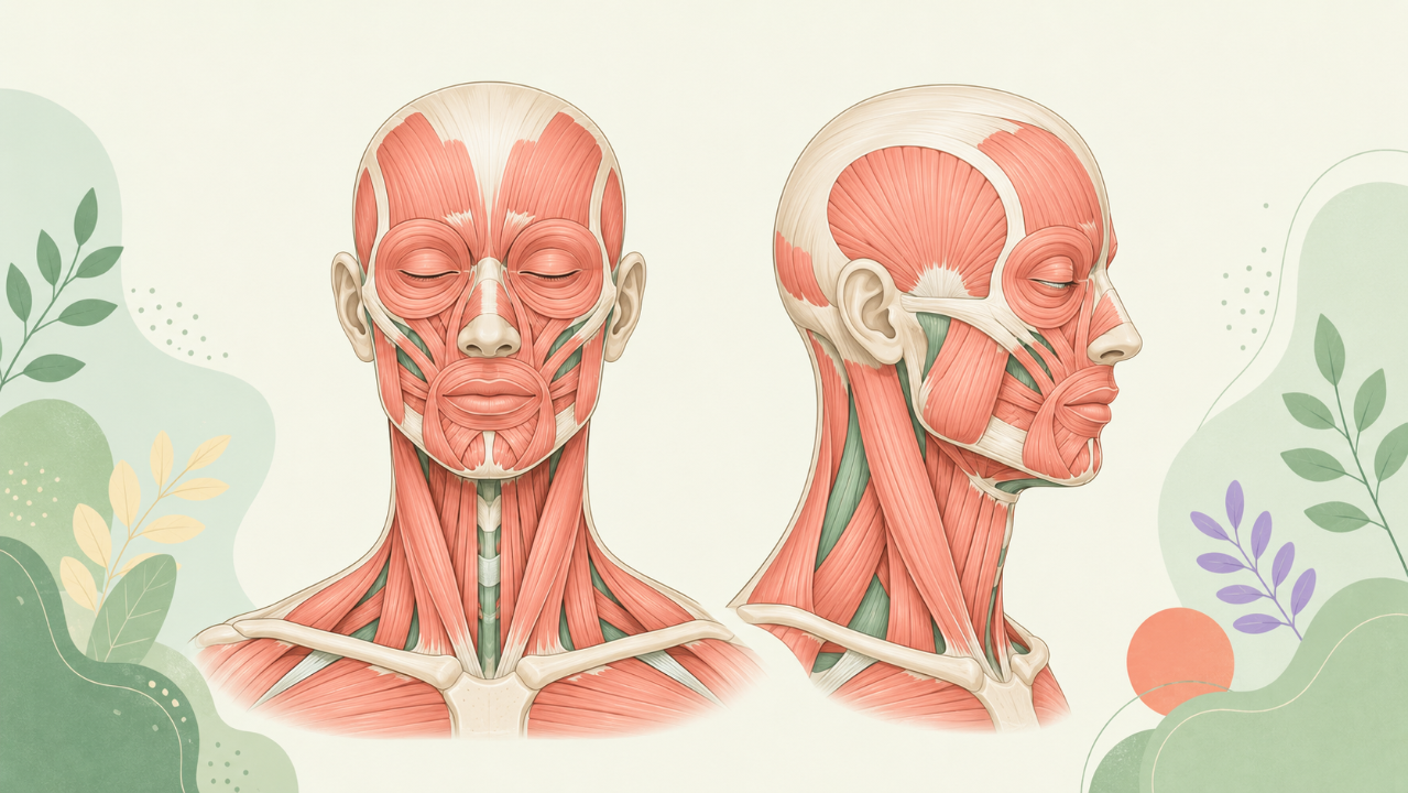
An Overview of Head and Neck Muscles
Classification of Head and Neck Muscles
Head and neck muscles can be classified according to their functions:
- Masticatory muscles (masseter, temporalis, medial and lateral pterygoids) responsible for biting and chewing actions
- Facial expression muscles (oculo-facial, orbicularis oculi, buccinator, orbicularis oris, levator labii superioris, zygomaticus major and minor, risorius, depressor anguli oris, mentalis) enabling facial expressions
- Laryngeal muscles (thyrohyoid, stylopharyngeus, posterior cricoarytenoideus, lateral cricoarytenoideus, transverse and oblique arytenoideus, inferior constrictor pharyngis, superior and inferior longitudinal pharyngis, circopharyngeus) involved in swallowing, speech production, and maintaining airway patency
- Cervical muscles (sternocleidomastoid, omohyoideus, scalene, longus colli, rectus capitis anterior and posterior, obliquus capitis superior and inferior, splenius capitis, semispinalis capitis, trapezius) responsible for head and neck posture maintenance and movements
Muscle Structure
Muscles of the head and neck are composed of fibers bundled together to form fascicles, which in turn are arranged to form a muscle belly. Each muscle has a distinct origin and insertion point, defining its range of motion. The muscles' structure is further characterized by their tendons or aponeuroses at both ends, providing attachment sites.
Muscle Function
Muscles contract and relax through the action of motor neurons, which send signals via the peripheral nervous system. This process results in muscle fibers shortening, thereby causing movement or maintaining body posture. The specific function of each muscle can be determined by its origin and insertion points, as well as the direction of pull during contraction.
Masticatory Muscles
Masseter
Structure and Function
The masseter is a thick, flat muscle located on each side of the lower jaw. It originates from the zygomatic arch and inserts onto the angle of the mandible, acting to close the jaw during biting and chewing actions.
Temporalis
Structure and Function
The temporalis is a fan-shaped muscle located on the side of the skull, extending from the temporal bone anteriorly to the zygomatic arch posteriorly. It inserts onto the coronoid process and mandibular ramus of the mandible, participating in jaw opening and closing actions as well as lateral movements of the lower jaw.
Pterygoids
Structure and Function
The pterygoids (medial and lateral) are located on either side of the sphenomandibular ligament, within the temporomandibular joint. They work in conjunction with the masseter and temporalis muscles to facilitate jaw movements. The medial pterygoid assists in closing the jaw, while the lateral pterygoid aids in both opening and rotating the mandible.
Facial Expression Muscles
Orbicularis Oculi
Structure and Function
The orbicularis oculi encircles the eye, with origins on the bones surrounding the orbit (frontal bone, maxilla, zygomatic bone, and lacrimal bone) and insertions on the skin of the eyelids. It functions to close the eyelids, contributing to blinking and facial expressions.
Buccinator
Structure and Function
The buccinator is a thin, cylindrical muscle located deep within the cheek. It originates from the orbital part of the maxilla and zygomatic arch anteriorly and inserts onto the angular process of the mandible posteriorly. Its main function is to flatten the cheek during mastication, thus facilitating the movement of food into the back of the mouth.
Clinical Implications
Understanding the structure, function, and clinical significance of head and neck muscles is essential for various medical professionals, including dentists, oral surgeons, ENT specialists, and neurologists. Disorders affecting these muscles can result in impaired facial expressions, difficulty swallowing, pain, or even speech disorders. Early recognition and treatment of such conditions are crucial to ensure optimal patient outcomes.