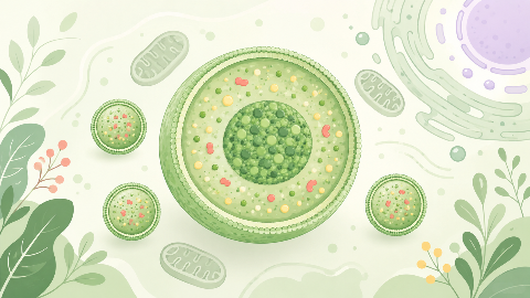
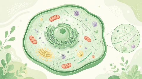
The peroxisomes
Discover peroxisomes, small cellular organelles that are key to the survival and adaptation of our cells! In this cell biology course, you'll explore their structure...
Osteology
In this osteology course, we explore the bony vomer, a key component of the mastoid and nasal anatomy of the skull. We will examine its role in essential functions such as chewing, breathing, and swallowing.
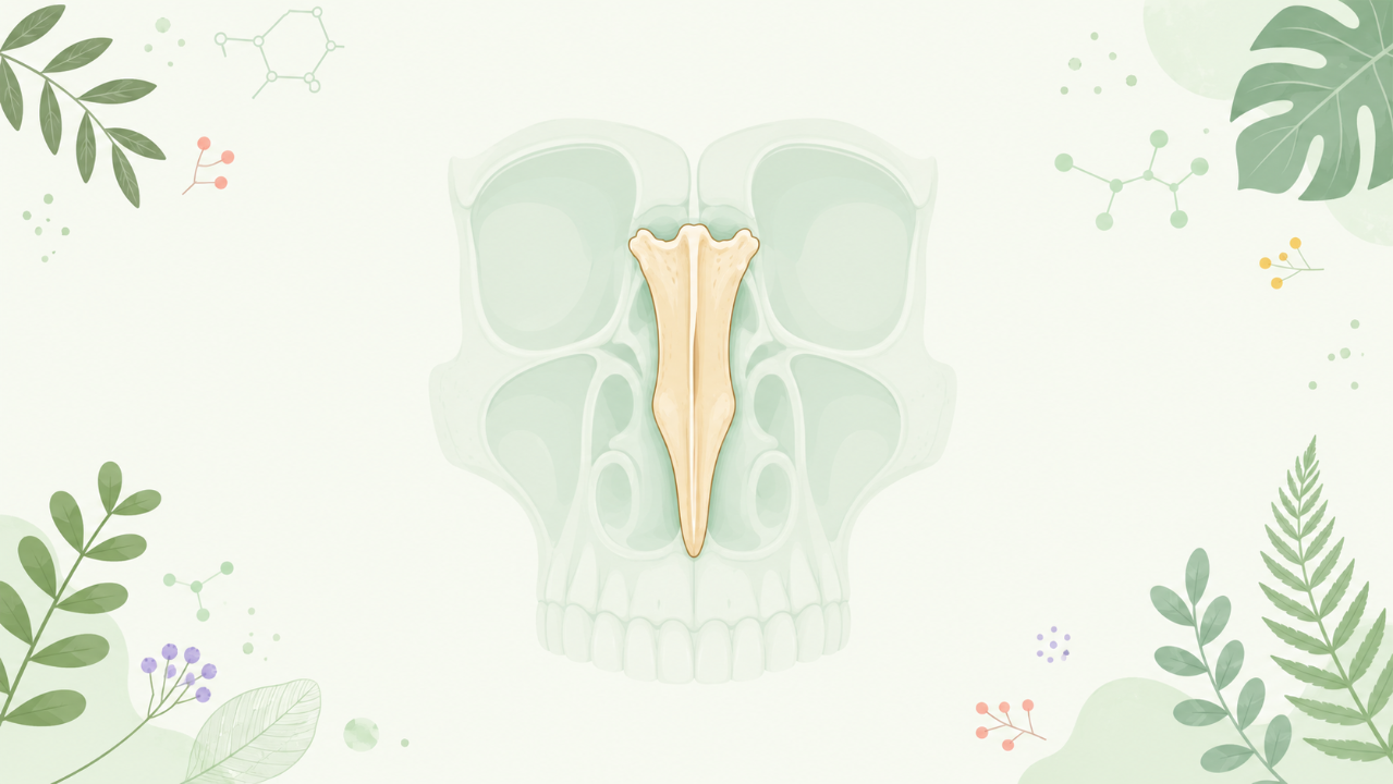
This comprehensive academic course provides a detailed exploration of the anatomical, evolutionary, and functional aspects of the vomer bone, a crucial element within the bony framework of the vertebrate nasal cavity. The vomer's significance is underscored by its role in supporting the roof of the mouth, articulating with other facial bones, and contributing to the nasal respiratory system. This course aims to equip students with a thorough understanding of the vomer bone's anatomy, development, variations, clinical relevance, and historical context.
The vomer bone has been recognized since antiquity as a distinctive feature of the vertebrate skull. Greek anatomists such as Herophilus (335–280 BC) and Erasistratus (304–250 BC) provided some of the earliest descriptions of this bone. Their observations were further built upon by Arabic scholars, including Ibn Sina (980–1037 AD), who contributed significantly to the understanding of human anatomy during the Middle Ages.
In contemporary osteological terminology, the vomer bone is classically considered one of the palatine bones, specifically the palatinum mediale. It is located in the midline of the hard palate, separating the two nasal cavities. Its name derives from the Latin word "vomere," meaning a plow or tongue-like instrument used for scraping or cleaning.
The vomer is a flat, plate-like bone that exhibits an asymmetrical shape, with a wider posterior portion and a narrower anterior section. Its rostral (front) end extends as a thin lamina that forms the anterior nasal spine, whereas its caudal (rear) extremity blends with the horizontal plates of the palatine bones to form the pterygoid hamulus.
The vomer articulates with several other bones in the skull, playing a crucial role in supporting the roof of the mouth and facilitating various movements. Its primary articulations include:
The vomer bone develops from the maxillary process of the medial nasal prominence during embryonic development. This process involves the fusion of several ossification centers, resulting in a fully formed adult vomer around the fifth fetal month.
Although rare, variations in the shape, size, or number of vomer bones can occur. Some of these anomalies may be linked to genetic factors or developmental abnormalities. Additionally, certain species have evolved unique adaptations related to their diet, environment, or reproductive strategies that influence the morphology and function of the vomer bone.
The vomer bone may be involved in various traumatic injuries, fractures, or developmental abnormalities that impact its function and overall health. Some common clinical conditions associated with the vomer include:
In forensic contexts, the examination of skull remains can provide valuable clues for identifying individuals based on the morphology, size, or degree of fusion of the vomer bone. These characteristics can aid in estimating age, sex, and racial affinities, thus contributing to the identification process during criminal investigations.
The vomer bone has undergone various modifications throughout vertebrate evolution, reflecting adaptations to different lifestyles, diets, and environments. For example, in certain aquatic species, the vomer may possess elongated processes for supporting the gill covers or aiding in suction feeding.
Comparative studies of the vomer bone across various vertebrate groups can provide insights into evolutionary trends and functional adaptations. For instance, in avian species, the vomer forms a significant part of the beak, while in reptiles, it contributes to the structure of the palate.
Do you think you know everything about this course? Don't fall into the traps, train with quizzes! eBiologie has hundreds of questions to help you master this subject.
Discover peroxisomes, small cellular organelles that are key to the survival and adaptation of our cells! In this cell biology course, you'll explore their structure...
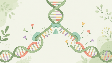
Discover how our DNA replicates with each cell division in this molecular biochemistry course: "DNA Replication." You'll learn the key steps in this crucial process...
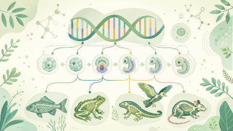
Learn about evolutionary developmental biology, the field that studies the mechanisms of embryonic development and their evolution at the molecular, cellular, and st...
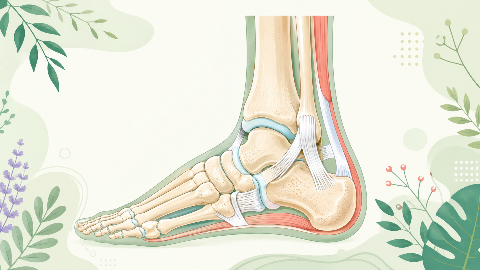
Discover the talonipedal joint in this syndesmology course: the bones, cartilage, and ligaments that enable the articulation of this complex joint. Learn to identify...
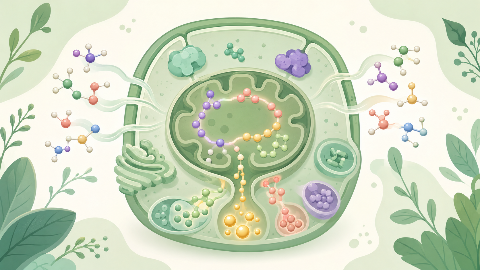
Discover amino acid metabolism in this exciting and technical course! You'll learn to identify the different types of amino acids and understand their essential role...
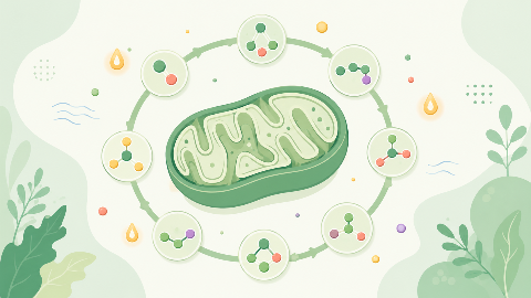
Discover the Krebs cycle: the central metabolic process that converts fatty acids into energy for the cell. Understand the steps in this process and learn how it int...
Dive into the intriguing world of Cellular Compartments! Explore the cytosol's structure, function, and dynamics, including its role in protein synthesis, energy pro...
