Osteology
course-show.h1-title
The sphenoid bone: a key bone for craniofacial balance and respiratory function. This study will immerse you in a deeper exploration of this complex bone, essential to the structure of the face and skull. You will learn to identify its anatomical features and understand its vital role in the muscular, nervous, and vascular systems.
Introduction
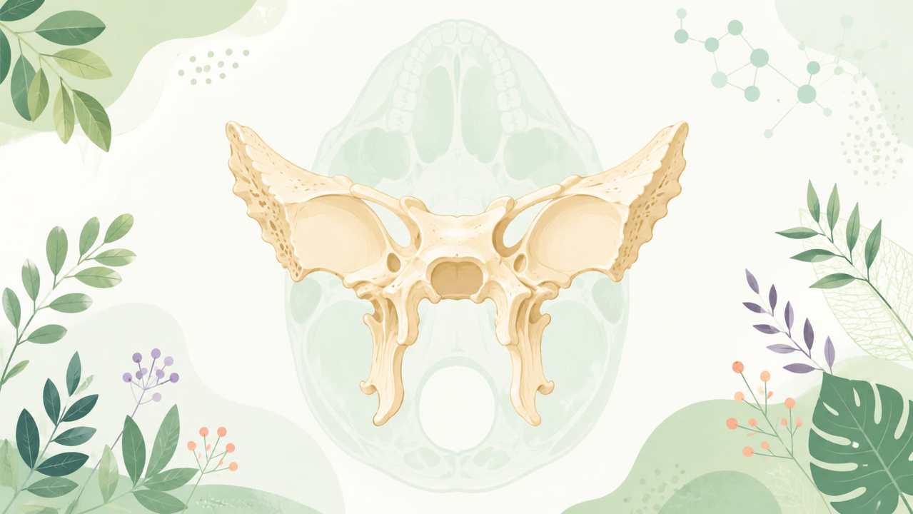
The sphenoid bone (or pterygo-palatine bone) is one of the cranial bones, located at the base of the skull and playing a crucial role in the structure and function of the head and facial region. This bone, shaped like a butterfly or bat wing, exhibits an intricate morphology, characterized by complex shapes and diverse components. The present academic course aims to offer an in-depth exploration of this bone's essential features, functions, development, and clinical relevance within the broader context of osteology.
Anatomy and Morphology
Overview
The sphenoid bone can be divided into four main parts: the greater wing (ala major), the lesser wing (ala minor), the body (corpus), and the sella turcica. Each part has unique characteristics and specific functions.
The Greater Wing
The greater wing is the largest and most lateral of the sphenoid's parts. It forms a significant portion of the temporal bone, contributing to the zygomatico-temporal suture with the zygomatic arch. Anatomically, it consists of two processes: the sphenopalatine (or posterior) and the zygomatic (or anterior).
Sphenopalatine Process
The sphenopalatine process is a curved projection that extends medially from the greater wing. It forms part of the nasal cavity's lateral wall, along with the maxillary bone and the palatine bone. It is also the site for the origin of the levator palpebrae superioris muscle, which elevates the upper eyelid.
Zygomatic Process
The zygomatic process projects anteriorly from the greater wing and articulates with the frontal bone to form part of the orbital rim. It also contributes to the zygomatico-maxillary suture, connecting the greater wing to the maxilla.
The Lesser Wing
The lesser wing is a smaller, more medial component of the sphenoid bone. It extends anterolaterally and articulates with the frontal bone to form part of the orbital floor. Anatomically, it consists of two processes: the frontal (or anterior) and the palatine (or posterior).
Frontal Process
The frontal process projects anteroinferiorly from the lesser wing and articulates with the frontal bone to form part of the orbital floor. It also contributes to the nasolacrimal groove, which drains tears into the nasal cavity.
Palatine Process
The palatine process projects posteriorly from the lesser wing and is continuous with the body of the sphenoid bone. It forms part of the hard palate, along with the palatine bone, and contributes to the pterygopalatine fossa, which houses various important structures.
The Body
The body is the central component of the sphenoid bone, located between the greater and lesser wings. It has a concave lateral surface, known as the convexity, that articulates with the temporal bone. Anatomically, the body can be divided into three regions: the anterior, middle, and posterior.
Anterior Region
The anterior region is the most lateral part of the body and forms part of the lateral wall of the orbit. It contributes to the infraorbital foramen, which transmits the infraorbital nerve and vessels.
Middle Region
The middle region is a transverse ridge that separates the anterior and posterior regions. It forms part of the roof of the pharynx (nasopharyngeal roof) and articulates with the basioccipital bone.
Posterior Region
The posterior region forms the roof of the nasopharynx and is continuous with the sella turcica. It houses the pituitary gland, an endocrine organ responsible for the production of various hormones essential for homeostasis and growth.
The Sella Turcica
The sella turcica is a small, saddle-shaped depression on the posterior region of the body, housing the pituitary gland (hypophysis cerebri). It has two parts: the anterior and posterior clinoids.
Anterior Clinoid Processes
The anterior clinoid processes are thin projections that extend anteromedially from the anterior part of the sella turcica. They form the medial walls of the cavernous sinuses, which contain important cranial nerves (III, IV, V1, and VI).
Posterior Clinoid Processes
The posterior clinoid processes are small projections that extend posterolaterally from the posterior part of the sella turcica. They articulate with the tentorium cerebelli, helping to maintain the position of the brainstem within the skull.
Development and Ossification
Embryonic Development
The sphenoid bone develops from five cartilages during embryogenesis: the alar, basisphenoid, palatine, quadratum, and vomer (which becomes part of the nasal cavity). These cartilages ossify at different stages, eventually forming the mature sphenoid bone.
Ossification
The ossification process begins in the embryonic stage and continues throughout fetal development and postnatal life. The alar cartilage gives rise to the greater wing (ala major), while the basisphenoid cartilage contributes to the body, sella turcica, and some parts of the lesser wing (ala minor). The palatine cartilage forms the palatine process of both wings, and the quadratum cartilage is incorporated into the body.
The ossification of the sphenoid bone is complex due to its intricate morphology. The different centers of ossification appear at various stages:
- Primary centers of ossification form in the greater wing and body during fetal life, fusing during the first few years of postnatal life.
- Secondary centers of ossification arise in the lesser wing, sella turcica, and basisphenoid during infancy and early childhood, fusing with the primary centers during adolescence.
- The sella turcica's anterior clinoid processes do not ossify until later in life, usually around age 20.
Clinical Relevance
Understanding the anatomy and development of the sphenoid bone is crucial for various medical specialties. For instance, neurosurgeons must be aware of the delicate structures housed within the sella turcica, such as the pituitary gland, to perform successful surgeries without causing complications. Orthodontists may also consider the position and development of the sphenoid bone when planning treatments for misaligned teeth or jaws.