Osteology
course-show.h1-title
Discover the world of feet: pedal osteology! Let's delve deeper into the bones of the human foot in this intensive course. We'll study the key anatomical structures in detail, understand their function, and discover how they relate to movement.
Introduction
This comprehensive and academically structured course aims to provide an in-depth understanding of foot osteology, a crucial aspect of anthropological and biological studies. The focus is on the skeletal elements that constitute the human foot and their functional roles.
Importance of Foot Osteology
Understanding foot osteology is essential for various fields including forensic science, paleontology, anthropology, and human evolution studies. It allows researchers to identify, compare, and analyze human skeletal remains, providing valuable insights into locomotion, adaptations, population migrations, and even disease patterns.
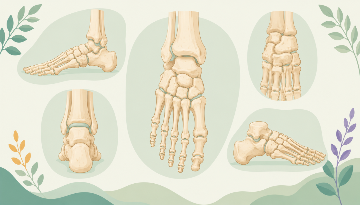
Skeletal Anatomy of the Foot
The Osseous Structures
The foot is composed of 26 bones: seven tarsals, five metatarsals, and fourteen phalanges (four distal phalanges for each foot). Each bone plays a unique role in supporting weight, propelling, and providing flexibility during locomotion.
Articulations
The joints of the foot enable mobility while maintaining structural integrity. The ankle joint (talocrural joint), tarsometatarsal joints, metatarsophalangeal joints, interphalangeal joints, and various synovial and non-synovial joints are crucial for proper foot function.
Bones of the Foot: Overview
Tarsal Bones
Talus: The talus is a wedge-shaped bone that articulates with the calcaneus, navicular, and cuboid bones, forming the ankle joint and tarsal bones complex. Its unique shape facilitates the foot's adaptation to different phases of gait.
Calcaneus: The calcaneus (heel bone) is the largest tarsal bone. It provides stability during weight-bearing and acts as a lever for propulsion in walking and running.
Navicular: The navicular bone connects the medial cuneiform and the talus, forming the arch of the foot. Its unique shape allows it to function as a shock absorber during impact.
Cuboid: The cuboid bone is located laterally in the midfoot, connecting the calcaneus with the metatarsal bones. It plays a crucial role in supporting the medial and lateral longitudinal arches of the foot.
Medial and Lateral Cuneiforms: These two irregularly shaped bones make up the medial and lateral columns of the midfoot, respectively. They contribute to the arch formation and provide stability during weight-bearing.
Three Small Tarsals (Talonavicular, Naviculocuboid, and Calcaneocuboid): These small tarsal bones form important joints with other tarsal and metatarsal bones, providing flexibility and structural integrity to the foot.
Metatarsal Bones
The five metatarsal bones are long, slender bones that connect the tarsals to the phalanges. They play a significant role in weight-bearing, propulsion, and flexibility during locomotion.
Phalanges
The fourteen phalanges are the distal bones of the foot. They consist of four distal phalanges for each foot, two proximal phalanges, and one intermediate phalanx for the first toe (hallux). These bones enable fine-tuning of the foot's movements during walking and running.
Foot Functional Anatomy: Overview
Understanding the function of each bone in the foot is essential to comprehend the overall mechanics of human locomotion. The foot plays a crucial role in shock absorption, weight distribution, propulsion, and balance maintenance during gait.
Phases of Gait
- Heel Strike: During this initial phase, the heel hits the ground, absorbing impact forces through the calcaneus and talus.
- Foot Flat Phase: The foot flattens, distributing weight evenly along the sole during mid-stance.
- Toe-off: As the body moves forward, the metatarsophalangeal joints flex, propelling the body forward with each step.
- Swing Phase: The leg swings forward, preparing for the next heel strike.
Conclusion
Understanding foot osteology provides invaluable insights into human evolution, locomotion adaptations, and various biological and anthropological studies. By studying the bones of the foot, their articulations, and functions, we can gain a deeper appreciation for this complex and essential structure.