Osteology
course-show.h1-title
Discover the secrets of the radius, a key bone in our human anatomy! With its complex structure and important mechanical functions, the radius is the foundation of our mobility and daily management.
Introduction
The human skeletal system is composed of 206 bones, one of which is the radius. This bone, located in the forearm, plays a crucial role in the articulation with the humerus (the upper arm bone) and the carpal bones of the wrist. The radius is an essential component of the lever system that enables various hand movements, such as gripping objects or rotating the forearm.
Anatomical Description
Gross Anatomy
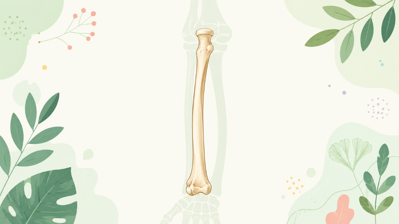
The human radius extends from the elbow to the wrist, with a slightly curved shape and a length of approximately 15 cm in an adult individual. It articulates with three bones: the humerus, the ulna, and several carpal bones (the scaphoid, lunate, triquetral, pisiform, and trapezium) of the wrist. The proximal end of the radius is cylindrical and articulates with the distal end of the humerus. Conversely, the distal end has a semilunar shape and forms joints with multiple carpal bones, primarily the scaphoid and lunate.
Histological Description
Like other bones in the human body, the radius is composed mainly of compact and spongy bone tissue. The compact bone forms the outer layer of the bone, providing strength and protection. It consists of tightly packed osteons (osteocytes within concentrically arranged lamellae) interconnected by canaliculi that facilitate nutrient exchange. On the other hand, the internal structure of the radius is made up of spongy or cancellous bone tissue, which contains numerous marrow cavities and is less dense than compact bone.
Functional Anatomy
Articular Surfaces and Movements
The proximal end of the radius has two articular surfaces: a smaller lateral surface that articulates with the radial notch (ulna) and a larger medial surface that articulates with the head of the humerus. These articulations enable flexion, extension, supination, and pronation movements at the elbow joint.
The distal end of the radius has a semilunar shape, with the lunate facet (located medially) forming an articular surface with the lunate bone and the radial tubercle (laterally) articulating with the scaphoid bone. These articulations allow for wrist movements such as flexion and extension, radial deviation (extension of the thumb side), and ulnar deviation (extension of the little finger side).
Muscles and Tendons
The radius is surrounded by muscles that originate from its surface or attach to it via tendons. The most important muscles acting on the radius include:
- Biceps brachii: Originates at the scapula and attaches to the radial tuberosity, flexing the elbow joint and supinating the forearm.
- Brachialis: Originates from the inferior surface of the humerus and inserts on the ulnar border of the radius, also flexing the elbow joint.
- Supinator: Originates at the posterior part of the scapula and attaches to the superior border of the radius, responsible for supinating the forearm.
- Pronator teres: Originates from the medial epicondyle of the humerus and inserts on the ulnar surface of the radius, pronating the forearm.
- Flexor carpi radialis: Originates at the medial supracondylar ridge of the humerus and attaches to the base of the second metacarpal bone, flexing the wrist joint and radial deviating it.
- Extensor carpi radialis longus: Originates from the lateral condyle of the humerus and attaches to the base of the third metacarpal bone, extending the wrist joint and radial deviating it.
Clinical Relevance
Due to its important role in hand movements and forearm articulations, injuries or diseases affecting the radius can lead to significant functional impairments. Common clinical conditions associated with the radius include:
- Fractures: These may occur due to traumatic events such as falls or direct blows to the forearm. Symptoms typically include pain, swelling, and difficulty using the affected arm. Treatment options depend on the severity of the fracture and may range from casting and immobilization to surgical intervention.
- Radial head subluxation: This condition involves partial or complete dislocation of the radial head from the annular ligament (a fibrous structure surrounding the proximal end of the radius). It can result from chronic repetitive motion injuries, such as those that occur during weight lifting or tennis playing. Symptoms include pain, instability, and difficulty rotating the forearm. Treatment may involve rest, immobilization, and physical therapy to restore proper function.
- Osteoarthritis: A degenerative joint disease characterized by the progressive wear and tear of articular cartilage. In the case of the radius, osteoarthritis can cause pain, stiffness, and limited mobility in the elbow or wrist joints. Treatment options include pain management, physical therapy, and joint replacement surgery in severe cases.