Angiology, vascular medicine or vascular medicine.
course-show.h1-title
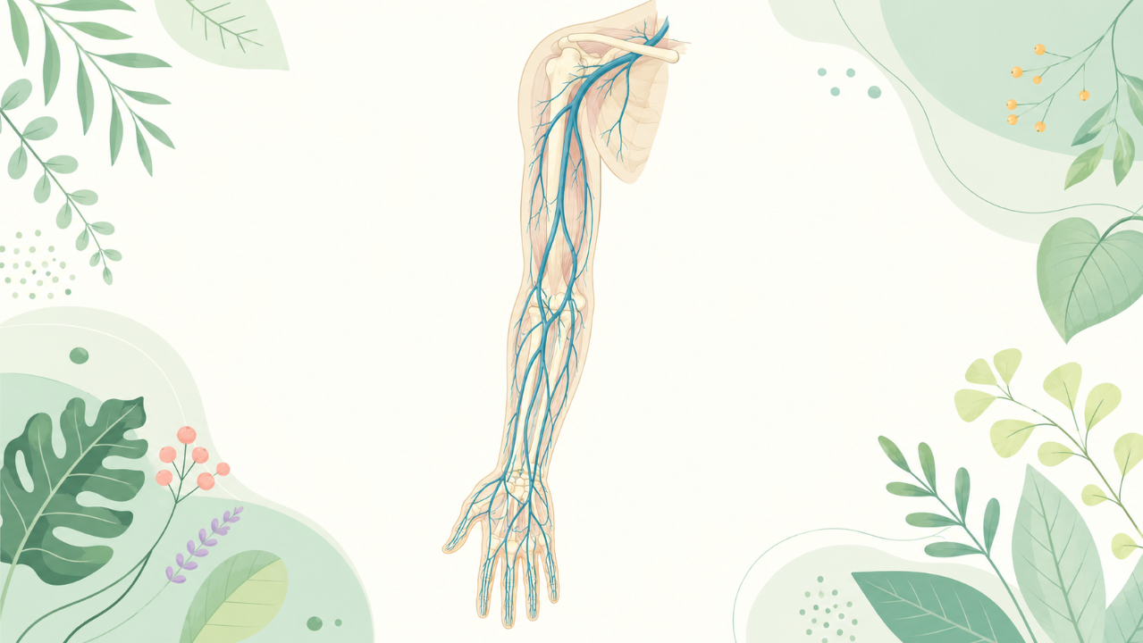
Discover the specific vascular anatomy of the human upper limb in this course. By exploring the vascular systems of the forearm and shoulders, you'll understand the morphology, function, and specific features of these vital structures. You'll also learn to identify common vascular anomalies in this anatomical region.
Introduction
The upper limb, comprising the arm and the forearm, is a complex anatomical region that houses numerous vessels. Understanding the structure and function of these vessels is essential for medical professionals involved in angiology, angiography, and interventional radiology, as well as surgical procedures related to the upper limb. This comprehensive course provides an in-depth analysis of the veins of the upper limb, focusing on their anatomy, physiology, clinical relevance, and diagnostic considerations.
Anatomical Overview of the Upper Limb
Bony Structure and Articulations
The upper limb is composed of three bones: the humerus, radius, and ulna. The articulation between these bones facilitates movements such as flexion, extension, supination, and pronation.
Soft Tissues and Nervous System
The muscles, tendons, ligaments, nerves, and blood vessels are encased within the soft tissues of the upper limb. The primary nerve supply is provided by the brachial plexus, which originates from the cervical spinal cord (C5-T1).
Venous Anatomy of the Upper Limb
Superficial Veins
Cephalic and Basilic Veins
The cephalic vein is a superficial vein that originates from the dorsal venous network of the hand. It courses up the anterior aspect of the arm, joining with the axillary vein at the lateral border of the pectoralis major muscle. The basilic vein begins in the palmar arch and travels along the medial side of the arm, ultimately emptying into the axillary vein near the medial border of the biceps brachii.
Other Superficial Veins
Several other superficial veins are present on the dorsum of the hand and forearm, including the dorsal metacarpal and digital veins, as well as the cephalic arch and the basilic arch. These veins play a role in the venous drainage of the upper limb but are less significant than the cephalic and basilic veins.
Deep Veins
Brachial, Radial, and Ulnar Veins
The deep veins of the upper limb are situated within the muscular compartments. The brachial vein arises from the union of the profunda brachii vein and the radial vein. It courses inferiorly along with the brachial artery, receiving tributaries such as the ulnar vein and numerous muscular veins. The radial vein originates at the wrist level from the junction of the dorsal venous network and the deep palmar arch. The ulnar vein begins at the level of the wrist joint, accompanying the ulnar artery up the forearm.
Muscle Venous Networks
The muscular compartments of the upper limb are drained by an extensive venous network. These veins connect with the brachial, radial, and ulnar veins to form a confluence of deep veins in the antecubital fossa.
Clinical Relevance and Diagnostic Considerations
Venous Insufficiency
Venous insufficiency is a common condition affecting the upper limb veins, particularly in the superficial venous system. Symptoms include edema, discomfort, and skin changes (e.g., hyperpigmentation, lipodermatosclerosis). Venous ulcerations can also occur in severe cases.
Thromboembolic Disease
Thrombotic events in the upper limb veins are relatively rare compared to those occurring in the lower limbs. Deep vein thrombosis (DVT) may result from trauma, surgery, or underlying conditions such as malignancy or hypercoagulability. Thromboembolic complications include pulmonary embolism and post-thrombotic syndrome.
Diagnostic Imaging Modalities
Various imaging techniques are used to diagnose and evaluate venous disorders in the upper limb, including:
- Duplex ultrasound: A non-invasive method that utilizes high-frequency sound waves to visualize veins and detect blood flow abnormalities.
- Venography: An invasive technique involving the injection of contrast medium into a vein, followed by radiographic imaging to assess venous anatomy and function.
- Computed tomography (CT) angiography: A non-invasive method that uses CT scanning to create detailed images of the blood vessels after the injection of a contrast agent.
- Magnetic resonance imaging (MRI): A non-invasive technique using magnetic fields and radio waves to produce detailed images of soft tissues, including veins.
Conclusion
The upper limb houses an intricate network of veins that play essential roles in the circulation of blood. Understanding their structure, function, and clinical relevance is crucial for medical professionals involved in the care of patients with vascular disorders. This comprehensive course aims to provide a detailed overview of the upper limb's venous system, encompassing its anatomy, physiology, and clinical implications.