Macroscopic anatomy
course-show.h1-title
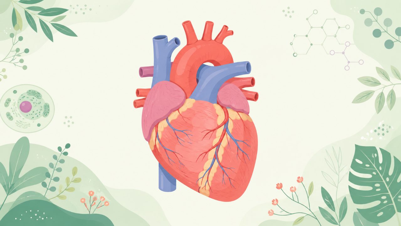
Discover the heart, the central organ that ensures blood circulation and exchanges between blood and the body. In this macroscopic anatomy course, we will explore its complex structure, function, and relationships with other organs. Learn to identify its different compartments, understand the blood vessels that circulate within it, and describe the most common heart diseases.
Introduction
The heart, a vital organ of the cardiovascular system, plays an essential role in maintaining life by pumping blood throughout the body to supply oxygen and nutrients to the tissues and organs. This comprehensive study aims to provide an in-depth analysis of the macroscopic structure, function, and physiology of the heart.
Overview
The heart is located in the mediastinum, a space within the thorax that separates the lungs. It has four chambers: two atria and two ventricles. The upper chambers, or atria, receive blood from various veins, while the lower chambers, or ventricles, pump the blood out to the rest of the body. The heart's structure is designed to facilitate efficient blood circulation, allowing for the continuous supply of oxygenated blood to the tissues and organs.
Macroscopic Anatomy
Gross Appearance
The heart has a triangular shape when viewed from the front or back and has a somewhat rectangular appearance when viewed from the sides. Its average weight is approximately 300 grams in adults, with dimensions of about 12-13 cm in length, 8-9 cm in width, and 6-7 cm in thickness.
Chambers
Right Atrium (Dextroatrium)
The right atrium receives deoxygenated blood from two large veins: the superior vena cava and the inferior vena cava. The wall of the right atrium is thin compared to the left atrium due to the lower pressure within this chamber.
Left Atrium (Sinistral Atrium)
The left atrium receives oxygenated blood from the pulmonary veins, which carry blood from the lungs. The left atrium's wall is thicker than that of the right atrium due to the higher pressure within this chamber.
Right Ventricle (Dextroventriculus)
The right ventricle pumps deoxygenated blood to the lungs via the pulmonary artery. The right ventricle's wall is relatively thin but strong to withstand the low pressure within this chamber.
Left Ventricle (Sinistroventriculus)
The left ventricle is the largest and strongest chamber of the heart, as it must pump oxygenated blood throughout the body at high pressures. Its wall is thick and muscular, allowing for efficient contraction.
Valves
Valves are essential structures within the heart that prevent backflow of blood. There are four valves in total:
- Tricuspid Valve (Ventriculoatrial Valve): separates the right atrium from the right ventricle. It has three cusps, or flaps, and is semi-lunar in shape.
- Mitral Valve (Atrioventricular Valve): separates the left atrium from the left ventricle. It also has three cusps but is more complex in structure due to its anatomical position.
- Pulmonary Semilunar Valve: separates the right ventricle from the pulmonary artery. It has three semi-lunar cusps.
- Aortic Semilunar Valve: separates the left ventricle from the aorta. It also has three semi-lunar cusps.
Function and Physiology
The heart's primary function is to pump blood throughout the body, supplying oxygen and nutrients to the tissues and organs. This is achieved through a series of coordinated contractions known as the cardiac cycle. The sequence of events includes:
- Atrial Contraction (Diastole)
- The atria fill with blood.
- Ventricular Filling (Diastole)
- Blood flows from the atria into the ventricles due to the difference in pressure between the two chambers.
- Ventricular Contraction (Systole)
- Both ventricles contract simultaneously, forcing blood out of the heart and into the aorta and pulmonary artery.
- Atrial Relaxation (Diastole)
- The atria relax, allowing blood to flow from the veins into the atria.
By following this cycle, the heart pumps blood continuously, ensuring that oxygenated blood is supplied to the tissues and organs.
Clinical Relevance
Understanding the structure and function of the heart is essential in diagnosing and treating cardiovascular diseases, which are among the leading causes of death globally. Some common cardiovascular conditions include coronary artery disease, hypertension, valvular heart disease, and congenital heart defects. Early detection and appropriate management can significantly improve outcomes for affected individuals.
Conclusion
This study provides an in-depth analysis of the macroscopic structure, function, and physiology of the heart. By understanding the complex interplay between its various components, we gain valuable insights into the mechanisms that support life itself. This knowledge is instrumental in developing effective strategies for diagnosing and treating cardiovascular diseases, ultimately improving the health and well-being of countless individuals worldwide.