Anatomy
course-show.h1-title
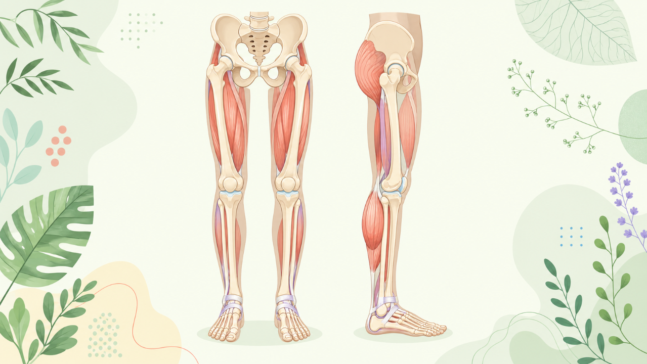
Discover the internal structures and how they work to understand the musculoskeletal system of the foot and thigh. Follow a clear presentation, enriched with illustrations, which will highlight the joints, muscles, nerves, and arteries. You will learn to identify and name these fundamental elements.
Introduction
The human lower limb, comprising the pelvis, hip, knee, and ankle regions, plays a crucial role in locomotion, weight-bearing, and postural support. This comprehensive guide aims to provide an academic and detailed exploration of the anatomical structures within the lower limb, essential for students pursuing advanced studies in Biology.
Overview of Lower Limb Regions
Pelvic Region
- Bones: The pelvis is composed of three bones: ilium, ischium, and pubis, which fuse during adulthood to form the innominate bone.
- Joints: The sacroiliac joint connects the pelvis with the sacrum, while the symphysis pubis is a cartilaginous joint between the pubic bones of the pelvis.
Hip Region
- Bones: The hip region primarily includes the coxal (pelvic) bone and the femur. The coxal bone consists of three fused bones: ilium, ischium, and pubis. The head of the femur forms the articulating surface of the ball-and-socket hip joint with the acetabulum, a cup-shaped cavity within the innominate bone.
- Joints: The primary joint in this region is the hip joint, which provides for flexion, extension, abduction, adduction, and circumduction of the lower limb.
Knee Region
- Bones: The tibia (shin bone) and fibula (calf bone) are the long bones in the leg, while the femur continues down to form the condyles of the knee joint.
- Joints: The knee region is dominated by the knee joint (tibiofemoral joint), a complex joint that enables flexion and extension of the lower limb. The patella (kneecap) protects the knee joint during movement.
Ankle Region
- Bones: The ankle region includes the tibia, fibula, talus, calcaneus, navicular bone, cuboid bone, and metatarsal bones.
- Joints: The primary joint in this region is the ankle joint (talocrural joint), which allows for plantar flexion, dorsiflexion, eversion, and inversion of the foot. Additionally, the subtalar joint and the inferior tibiofibrular joint are important for foot stability during movement.
Muscles and Fasciae
This section will delve into the numerous muscles and fasciae found within the lower limb, their origins, insertions, functions, and innervations.
Pelvic Region
- Muscles: The hip flexors, adductors, abductors, and rotators, along with the gluteal muscles.
- Fasciae: The iliotibial tract, the pelvic fascia, and the aponeurosis of the obturator internus muscle.
Hip Region
- Muscles: The hamstrings, adductors, quadriceps, and hip rotators.
- Fasciae: The iliofemoral ligament, the ischiofemoral ligament, and the joint capsule of the hip joint.
Knee Region
- Muscles: The muscles responsible for knee flexion, extension, and rotation, such as the quadriceps, hamstrings, gastrocnemius, and soleus.
- Fasciae: The patellar ligament, the joint capsule of the knee joint, and the anterior and posterior cruciate ligaments.
Ankle Region
- Muscles: The muscles responsible for plantar flexion, dorsiflexion, eversion, and inversion of the foot, such as the tibialis anterior, peroneus longus, and gastrocnemius.
- Fasciae: The plantar aponeurosis (plantar fascia) and the deep and superficial fibular compartments.
Nerves and Vessels
This section will examine the nerves and blood vessels that supply the lower limb, their origins, courses, branches, and functional significance.
Pelvic Region
- Nerves: The femoral nerve, obturator nerve, and iliohypogastric nerve.
- Vessels: The external iliac artery, internal iliac artery, and the femoral artery.
Hip Region
- Nerves: The sciatic nerve, the femoral nerve, and the obturator nerve.
- Vessels: The femoral artery and its branches, as well as the circumflex iliac vessels.
Knee Region
- Nerves: The sciatic nerve, tibial nerve, common peroneal nerve, and the saphenous nerve.
- Vessels: The popliteal artery, anterior tibial artery, posterior tibial artery, and the fibular (peroneal) artery.
Ankle Region
- Nerves: The deep peroneal nerve, superficial peroneal nerve, tibial nerve, and sural nerve.
- Vessels: The posterior tibial artery, dorsal pedis artery, and the plantar arch vessels.
Conclusion
This comprehensive guide aims to provide an in-depth understanding of the anatomy of the lower limb, essential for students pursuing advanced studies in Biology. The lower limb consists of four primary regions (pelvic, hip, knee, and ankle) with numerous bones, joints, muscles, fasciae, nerves, and vessels within each region. A thorough understanding of these structures will enable students to appreciate the complexity and intricacies of human locomotion and postural support systems.