Osteology
course-show.h1-title
Discover the secrets of the inguinal bone by exploring the world of osteology in our course "The Coxal Bone." By learning to identify and understand the structure, function, and anatomy of the different parts of this essential bone, you'll acquire the skills needed to analyze a multitude of clinical cases. You'll also benefit from an interdisciplinary approach to studying the evolution of this structure in nature.
Introduction
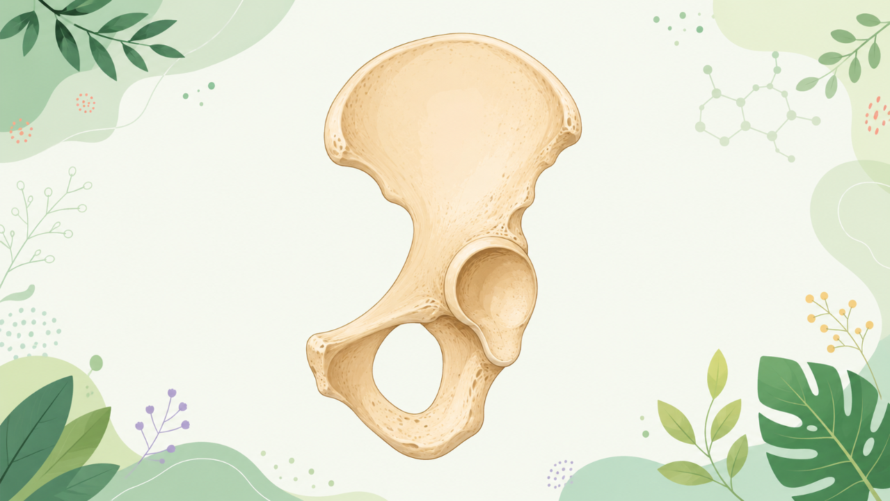
The hip bone, more formally known as the innominate or os coxae, is an essential component of the human skeleton. This bone serves multiple functions in locomotion and provides a site for attachment of various muscles. The hip bone consists of three fused bones: ilium, ischium, and pubis, which are united at their ventral ends by the acetabulum, a socket that articulates with the head of the femur to form the ball-and-socket joint. This tutorial aims to provide a comprehensive, detailed, and hierarchized overview of the hip bone's anatomy, structure, and functions within the context of osteology.
Bony Structure of the Hip Bone
Ilium
The ilium is the largest and uppermost portion of the hip bone, contributing significantly to the pelvic girdle's stability. The ilium is characterized by its broad, fan-shaped form, which consists of three prominent projections: the superior, anterior, and posterior surfaces. Each surface displays unique anatomical landmarks.
Superior Surface (Dorsal Surface)
The superior surface has several distinct features, including the following:
- Iliac Crest: The iliac crest is a ridged, horizontal ridge that extends along the lateral border of the superior surface. It provides attachment sites for various muscles, such as the gluteus maximus and tensor fascia lata.
- Iliac Tubercle: The iliac tubercle, located on the posterior aspect of the superior surface, serves as an origin site for the gluteus minimus and gluteus medius muscles.
- Spinal Articulation: The anterior and posterior superior iliac spines (ASIS and PSIS) are two prominent bony projections that mark the attachment sites for the thoracolumbar fascia, ligamentum sacrospinale, and various abdominal muscles. The ASIS lies anterior to the PSIS, forming a transverse ridge called the arcuate line.
Anterior Surface (Ventral Surface)
The anterior surface displays several significant anatomical features:
- Iliac Pubic Rami: The iliac pubic rami are two curved, blade-like processes that extend inferiorly and medially from the ventral surfaces of the ilium. They meet with the respective pubic rami to form the public symphysis.
- Anterior Superior Iliac Spine (ASIS): The ASIS lies at the most anterior and superior aspect of the anterior surface, forming part of the pelvic rim.
- Iliac Crest Articulation: The iliac crest continues onto the anterior surface as a ridge, where it connects with the iliopubic eminence and forms a small depression called the inguinal ligament's insertion site.
Posterior Surface (Dorsal Surface)
The posterior surface of the ilium displays several critical anatomical landmarks:
- Iliac Tubercle: The iliac tubercle, a prominent projection located on the posterior aspect of the superior surface, continues onto the posterior surface as well. It serves as the origin site for the gluteus minimus and gluteus medius muscles.
- Greater Sciatic Foramen: This foramen, situated posterolateral to the iliac tubercle, transmits the sciatic nerve and other vessels.
- Posterior Inferior Iliac Spine (PSIS): The PSIS lies at the most posterior aspect of the posterior surface, forming part of the pelvic rim.
Ischium
The ischium is the inferior and medial portion of the hip bone, extending from the ventral border of the ilium to the ischiopubic ramus. The ischium comprises three parts: the body, ramus, and tuberosity.
Body
The body of the ischium is a broad, flat, curved plate that extends inferiorly from the ventral border of the ilium. The body's posterior surface features the ischial spine and ischial tuberosity.
Ramus
The ramus of the ischium projects laterally from the body, forming the ischiopubic ramus with the pubis. It extends to the ischial tuberosity and provides attachment sites for various muscles, such as the gluteus maximus and piriformis.
Tuberosity
The ischial tuberosity is a prominent, rounded projection that extends inferiorly from the body and ramus. The tuberosity serves as the insertion site for several important muscles, including the hamstrings (biceps femoris, semimembranosus, and semitendinosus) and the gluteus maximus's lower fibers.
Pubis
The pubis is the anterior portion of the hip bone, located between the ilium and the ischium. It consists of two bones joined by the public symphysis: the right and left pubic bones. The pubis features three parts: the body, ramus, and symphysis.
Body
The body of the pubis is a flat, horizontally oriented plate that extends anteriorly from the ventral border of the ilium. The body's inferior surface forms part of the pelvic floor and provides attachment sites for several muscles, such as the adductor longus and gracilis.
Ramus
The ramus of the pubis projects medially from the body and meets with the respective ramus of the ischium to form the ischiopubic rami. The ramus's inferior surface features the obturator foramen, through which the obturator nerve, artery, and vein pass.
Symphysis
The symphysis is the region where the right and left pubic bones fuse, forming the public symphysis. This joint provides a site for attachment of various ligaments, such as the arcuate, interpubic, and pubocervical ligaments. The symphysis also contributes to the pelvic floor's stability.
Acetabulum
The acetabulum is a shallow, deepened portion of the hip bone that serves as the socket for the head of the femur in the ball-and-socket joint. The acetabulum is formed by the fusion of the ilium, ischium, and pubis at their ventral ends. The acetabulum's articular surface is covered with hyaline cartilage, providing a smooth surface for articulation. The acetabular labrum, a fibrocartilaginous rim surrounding the acetabulum's margin, deepens and strengthens the socket, enhancing joint stability.
Muscles Attached to the Hip Bone
Several muscles originate or insert on various surfaces of the hip bone, including the following:
- Gluteus Maximus: Origins from the ilium's iliac crest and inserts on the ischial tuberosity.
- Gluteus Medius: Originates from the ilium's iliac crest and inserts onto the greater trochanter of the femur.
- Gluteus Minimus: Origins from the ilium's iliac fossa and inserts onto the greater trochanter of the femur.
- Piriformis: Originates from the pelvic surface of the sacrum and inserts on the greater trochanter of the femur via the superior gemellus muscle.
- Obturator Internus: Originates from the inferior ramus of the pubis, obturator membrane, and ischial ramus and inserts onto the lesser trochanter of the femur.
- Adductor Longus: Originates from the anterior inferior iliac spine (AIIS) and inserts on the linea aspera of the femur.
- Gracilis: Originates from the anterior surface of the body of the pubis and inserts onto the medial condyle of the tibia.
- Tensor Fascia Lata: Originates from the iliac crest's anterior superior border and inserts onto the iliotibial tract on the lateral condyle of the tibia.
- Biceps Femoris: Originates from the ischial tuberosity and inserts onto the head of the fibula and the lateral condyle of the tibia.
- Semimembranosus: Originates from the ischial tuberosity and inserts onto the medial condyle of the tibia.
- Semitendinosus: Originates from the ischial tuberosity and inserts onto the medial condyle of the tibia.
Clinical Relevance
Understanding the hip bone's structure, function, and muscular attachments is essential for various clinical applications. Common pathologies affecting the hip include osteoarthritis, fractures, dislocations, labral tears, and inflammatory diseases such as tendinitis or bursitis. A thorough knowledge of the hip bone's anatomy can aid healthcare professionals in diagnosing and treating these conditions effectively.
Conclusion
The hip bone plays a crucial role in human locomotion, providing both stability and mobility for the lower limbs. This tutorial has provided a comprehensive overview of the hip bone's structure, including its three constituent bones (ilium, ischium, and pubis), their distinct surfaces and landmarks, as well as the muscles that attach to this essential bone. Understanding the intricacies of the hip bone's anatomy can contribute significantly to clinical practice and promote effective diagnosis and treatment of various conditions affecting this region.