Osteology
course-show.h1-title
Join us for an immersion into the osteology of the lower limbs! Discover the complex structure of the leg and foot bones, and learn to identify the distinctive characteristics of each element.
Introduction
The study of osteology focuses on the bones and their structures, development, and evolution in organisms. This course delves into the osteology of the lower limb, providing a comprehensive understanding of the anatomy and functions of the bones that make up this region in human beings. The emphasis is on the detailed exploration of each bone, its morphology, and its role in the lower limb's overall structure and function.
Background and Importance
The lower limb, also known as the leg, is a crucial part of the human skeletal system. It consists of bones that support the body weight, provide mobility, and protect vital organs. Understanding the osteology of the lower limb is essential for medical professionals, anthropologists, and students of anatomy to gain insights into the intricacies of locomotion, evolutionary adaptations, and pathologies associated with this region.
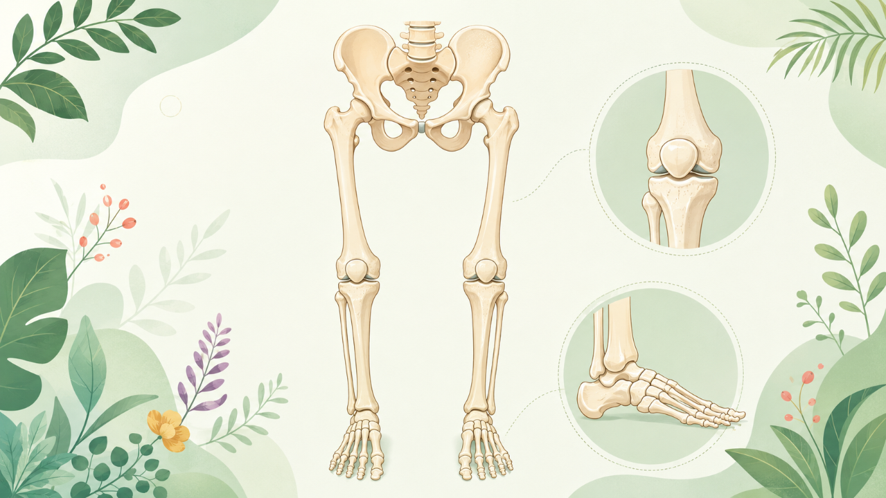
Lower Limb Anatomy
The lower limb can be divided into three regions: the hip region, the knee region, and the ankle region. Each region comprises several bones that work together to form a complex structure essential for various movements and functions.
Hip Region
The hip region is made up of the pelvis (hip bone) and the femur (thigh bone). The hip bone consists of three fused bones: ilium, ischium, and pubis. The femur is the longest and strongest bone in the human body, connecting the pelvis to the knee.
Ilium
The ilium forms the majority of the hip bone, making up its posterior and lateral portions. It has a broad, flattened articular surface for articulation with the sacrum and coccyx, forming the posterior pelvic inlet. The iliac crest, a prominent ridge on the posterior superior aspect of the ilium, serves as an attachment site for various muscles.
Ischium
The ischium forms the inferior and medial part of the hip bone. It has two significant projections: the ischial tuberosity and the ischial spine. The ischial tuberosity is a large, roughly triangular bony prominence that serves as an attachment site for several muscles, tendons, and ligaments.
Pubis
The pubis forms the anterior and medial part of the hip bone. It has two rami (rami superior et rami inferior) that meet at a symphysis pubis, a cartilaginous joint in the midline. The pubic rami serve as attachment sites for various muscles and ligaments.
Femur
The femur is a long, heavy bone with a round head at its upper end that articulates with the acetabulum of the hip bone. The shaft of the femur is strong and cylindrical, with two condyles on its lower end that articulate with the tibia and fibula in the knee joint.
Knee Region
The knee region consists of the femur (from the hip), tibia (shinbone), fibula (calculus lateralis), and patella (kneecap). The major joint in this region is the knee joint, where the femur articulates with the tibia.
Tibia
The tibia is the larger and more medial of the two bones forming the lower leg. It has a broad articular surface on its upper end that articulates with the femur to form the knee joint. The lower end of the tibia has two condyles, one medial (tibial medial condyle) and one lateral (tibial lateral condyle), that articulate with the talus in the ankle joint.
Fibula
The fibula is the smaller and thinner bone of the lower leg, situated lateral to the tibia. It has a thin articular surface on its upper end for articulation with the tibia, forming the syndesmosis (a strong ligamentous joint). The distal end of the fibula is narrow and pointed, with a small process (malleolus fibularis) that forms part of the ankle joint.
Patella
The patella, or kneecap, is a sesamoid bone located at the front of the knee joint. It protects the knee's extensor mechanism and improves the lever arm for the quadriceps muscles during knee extension.
Ankle Region
The ankle region includes the talus (talus), calcaneus (heel bone), navicular (navicula), cuboid (cuboid bone), and cuneiforms (three small bones: lateral, intermediate, and medial). These bones form the tarsus, or ankle joint, which articulates with the distal end of the fibula and tibia.
Talus
The talus is a wedge-shaped bone situated between the tibia and fibula above and the calcaneus below. It has a convex articular surface on its superior surface for articulation with the tibia, fibula, and navicular. The posterior process of the talus forms part of the subtalar joint, which allows inversion and eversion movements at the ankle.
Calcaneus
The calcaneus is the largest bone in the foot, forming the heel. It has a large, concave articular surface on its inferior surface for articulation with the talus, cuboid, and cuneiforms. The posterior portion of the calcaneus forms the calcaneal tuberosity, which serves as an attachment site for various muscles and ligaments.
Navicular
The navicular is a large, irregularly shaped bone situated between the tibia and talus above and the cuboid below. It has a prominent medial process that articulates with the talus and a lateral process that articulates with the cuboid. The body of the navicular forms part of the arches of the foot.
Cuboid and Cuneiforms
The cuboid and cuneiforms are small bones situated lateral to the navicular, forming the lateral column of the midfoot. They have various articular surfaces that connect with each other and the calcaneus, talus, and metatarsals. The cuneiforms also contribute to the formation of the arches of the foot.