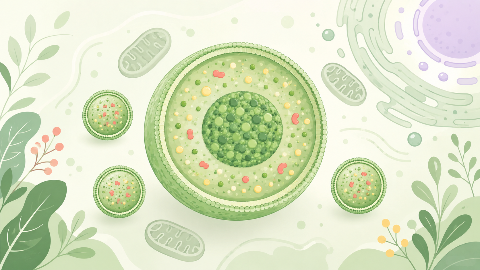
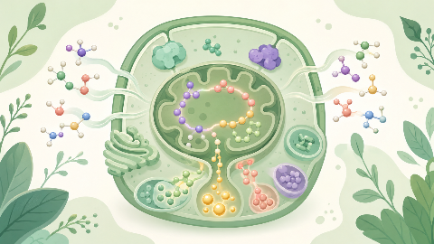
The peroxisomes
Discover peroxisomes, small cellular organelles that are key to the survival and adaptation of our cells! In this cell biology course, you'll explore their structure...
Myology
In this myology course, we explore the calf muscles, which play a vital role in our body's mobility and stability. We study their anatomy, function, and control, as well as their interactions with other muscle structures. With this in-depth knowledge of the calf muscles, we can better understand the origins and treatments of calf-related disorders.
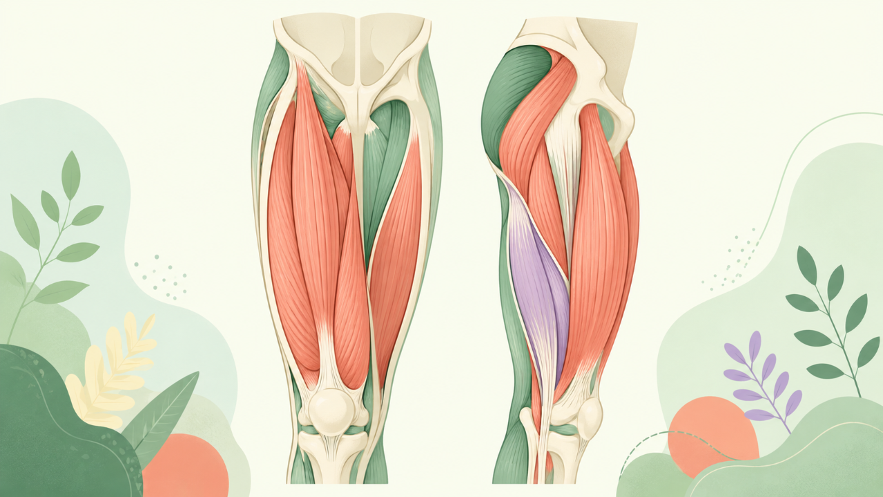
The human thigh is a vital region of the musculoskeletal system, housing several essential muscle groups that enable locomotion, stability, and postural control. This comprehensive course delves into an in-depth analysis of the muscles constituting the thigh, highlighting their functions, origins, insertions, actions, innervations, and relationships with other structures.
The human thigh is divided into three regions: anterior, posterior, and medial. Each region houses unique muscle groups responsible for different movements and functions.
The quadriceps femoris muscle group is primarily responsible for knee extension and stabilization during various movements, such as walking, running, and jumping. It consists of four muscles: rectus femoris, vastus lateralis, vastus intermedius, and vastus medialis. Each has distinct origins, insertions, and actions within the leg joints.
The hamstrings consist of three muscles - semitendinosus, semimembranosus, and biceps femoris. They primarily function in knee flexion (bending) and hip extension (straightening). Each muscle has distinct origins, insertions, and actions within the leg joints.
The popliteus muscle is essential for rotating the tibia during flexion, stabilizing the knee joint, and facilitating the transition from heel strike to mid-stance in locomotion. It originates from the lateral femoral condyle and inserts on the fibular head.
The adductor group is responsible for thigh adduction, medial rotation of the leg, and maintaining the pelvis' stability during various movements. They consist of six muscles - gracilis, pectineus, adductor longus, adductor brevis, adductor magnus (long head), and adductor magnus (short head).
Thigh muscles share close relationships with various structures within the leg joints, tendons, ligaments, and aponeuroses. Understanding these relationships is crucial for understanding their functions, movements, and potential injuries.
Understanding the anatomy, function, and relationships of thigh muscles is essential in diagnosing and treating related injuries, as well as developing effective exercise programs for rehabilitation or athletic performance enhancement. Common injuries affecting these muscles include strains, sprains, contusions, tendinitis, and tears.
This comprehensive course has explored the muscles of the thigh, highlighting their unique functions, origins, insertions, actions, innervations, and relationships with other structures. By gaining a deeper understanding of these essential muscle groups, students can better appreciate the complexity of human anatomy, the intricacies of the musculoskeletal system, and how to apply this knowledge in clinical practice or exercise programming.
Do you think you know everything about this course? Don't fall into the traps, train with quizzes! eBiologie has hundreds of questions to help you master this subject.
Discover peroxisomes, small cellular organelles that are key to the survival and adaptation of our cells! In this cell biology course, you'll explore their structure...
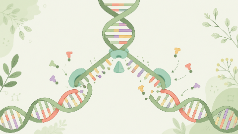
Discover how our DNA replicates with each cell division in this molecular biochemistry course: "DNA Replication." You'll learn the key steps in this crucial process...
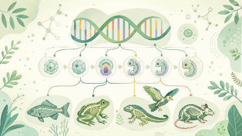
Learn about evolutionary developmental biology, the field that studies the mechanisms of embryonic development and their evolution at the molecular, cellular, and st...
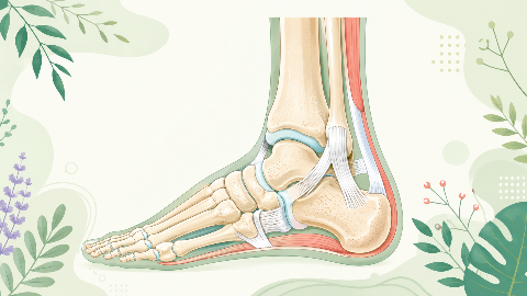
Discover the talonipedal joint in this syndesmology course: the bones, cartilage, and ligaments that enable the articulation of this complex joint. Learn to identify...
Discover amino acid metabolism in this exciting and technical course! You'll learn to identify the different types of amino acids and understand their essential role...
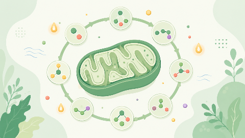
Discover the Krebs cycle: the central metabolic process that converts fatty acids into energy for the cell. Understand the steps in this process and learn how it int...
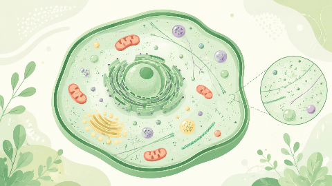
Dive into the intriguing world of Cellular Compartments! Explore the cytosol's structure, function, and dynamics, including its role in protein synthesis, energy pro...
