Osteology
course-show.h1-title
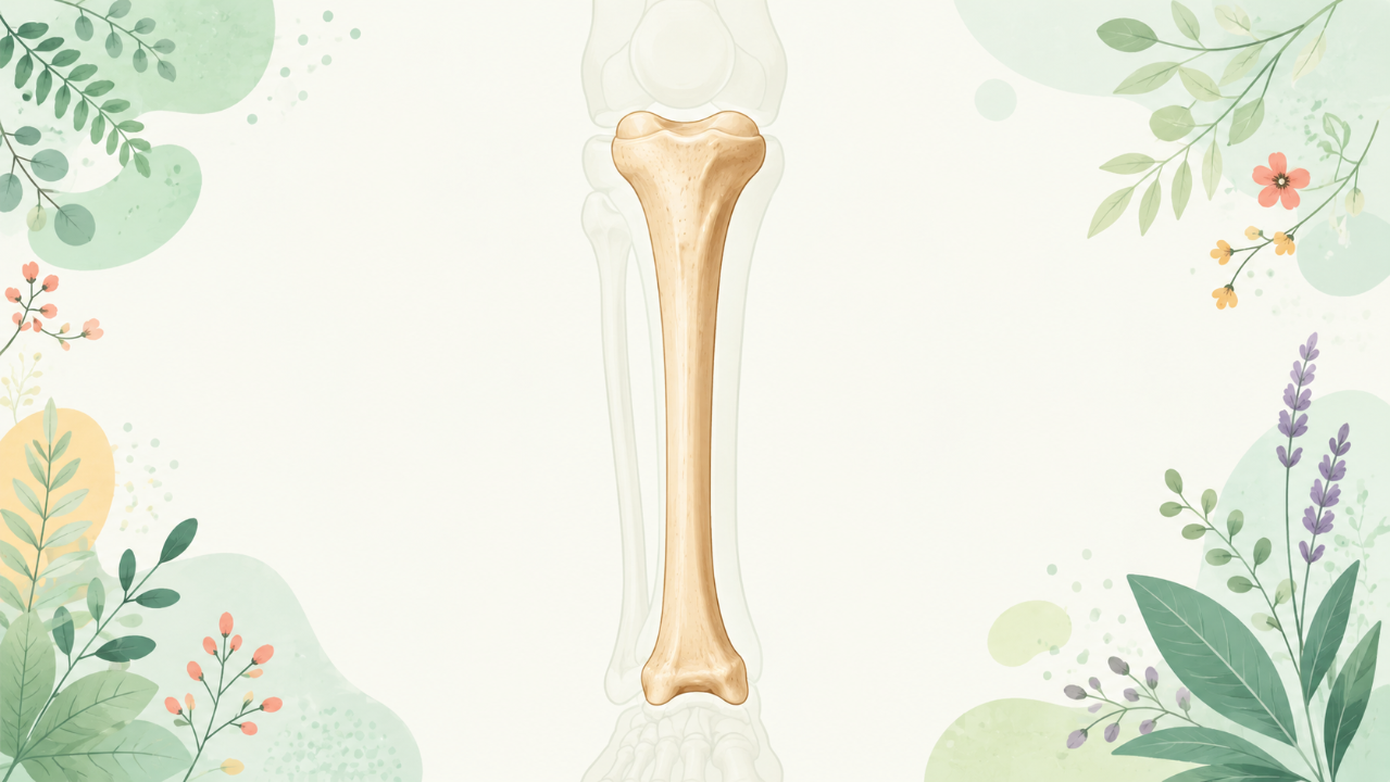
Discover the anatomy of the human ankle in our osteology course: "The Tibia." Learn to identify its structure, functions, and characteristics. Join us for a detailed exploration of the lower limb.
Introduction
The tibia, also known as the shinbone, is a crucial component of the human lower limb skeleton. It plays an integral role in articulation with other bones and provides essential support for various physical activities such as walking, running, and jumping. This comprehensive course will delve into the morphology, osteohistology, developmental biology, evolutionary history, clinical relevance, and comparative anatomy of the tibia.
Anatomical Position and Relationships
In the human skeleton, the tibia is located in the lower limb between the femur and fibula. The anterior surface faces forward, while the posterior surface faces backward. The medial side is positioned closer to the midline of the body, whereas the lateral side is further away.
Bony Relations
The tibia articulates with several bones in the lower limb:
- Femur: The proximal end of the tibia forms a synovial joint called the knee joint (tibiofemoral joint) with the distal end of the femur, allowing flexion and extension movements.
- Patella: The patella, or kneecap, is a sesamoid bone that protects the anterior knee and improves the efficiency of the quadriceps during leg extension.
- Fibula: The fibula articulates with the lateral condyle of the tibia at the ankle joint (tibiofibular syndesmosis) and forms the interosseous membrane that connects both bones.
- Talus: At the distal end, the tibia forms the medial malleolus, which articulates with the talus bone at the ankle joint (tibiotarsal joint), providing stability during weight-bearing activities.
Morphology and Osteohistology
The tibia can be divided into three regions: proximal, shaft, and distal. Each region has distinct characteristics in terms of shape and osteohistological composition.
Proximal Region
- Shape: The proximal end is expanded laterally to form the medial and lateral condyles. These condyles articulate with the femur at the knee joint, allowing for flexion, extension, and rotation of the lower limb.
- Osteohistology: The articular surface of the condyles is covered by hyaline cartilage to facilitate smooth movement between bones. The underlying bone contains a high percentage of spongy bone (cancellous bone) with numerous blood vessels and marrow spaces. The shaft of the tibia has a predominantly compact bone structure, providing strength and resistance to compression forces.
Shaft Region
- Shape: The shaft is long and cylindrical in shape, with slightly convex surfaces on both anterior and posterior aspects. This allows for rotation around its longitudinal axis during activities such as walking.
- Osteohistology: The shaft has a predominantly compact bone structure, consisting of osteons arranged parallel to the long axis of the bone. This provides resistance to compressive forces encountered during weight-bearing activities.
Distal Region
- Shape: The distal end is expanded medially to form the medial malleolus, which articulates with the talus at the ankle joint. The lateral aspect of the distal end is relatively flat and forms part of the ankle mortise.
- Osteohistology: Similar to the proximal region, the articular surface of the distal end is covered by hyaline cartilage to facilitate smooth movement at the ankle joint. The underlying bone contains a mix of compact and spongy bone, with numerous blood vessels and marrow spaces.
Developmental Biology and Evolutionary History
The tibia develops as part of the limb bud during embryonic development. The ossification process begins in the center of the diaphysis (shaft) and progresses toward both ends, eventually forming a fully ossified bone.
Throughout evolution, the tibia has undergone various modifications to adapt to different locomotive strategies. In primates, including humans, the tibia became more robust to accommodate upright posture and bipedal locomotion.
Clinical Relevance
The tibia is susceptible to fractures due to its role in weight-bearing activities. Common causes of tibial fractures include motor vehicle accidents, falls, and sports injuries. Other clinical conditions that may affect the tibia include osteomyelitis (bone infection) and fractures during growth periods leading to abnormal bone development (blount's disease).
Comparative Anatomy
While the overall structure of the tibia is conserved across vertebrates, there are variations in size, shape, and articulations due to differences in locomotive strategies. For example, in birds and quadrupedal mammals, the tibia plays a crucial role in weight-bearing and propulsion during running or jumping. In contrast, in some aquatic animals like whales and dolphins, the tibia is greatly reduced or absent due to their primary mode of locomotion being swimming rather than walking or running.