Osteology
course-show.h1-title
Discover the femur, a keystone of the human skeletal system. This osteology unit will allow you to explore the internal and external structures of this important bone. Also, decipher its role in locomotion, its varied morphology, and its connections with other lower limbs.
Introduction
In this comprehensive academic course, we delve into a detailed exploration of the femur, the longest bone in the human body. Our focus is on providing a thorough understanding of its anatomy, morphology, physiology, and clinical significance within the realm of osteology.
The Femur: An Overview
The femur, also known as the thigh bone, is a long, stout, and heavy bone that forms the upper part of the leg. It connects the hip to the knee joint and plays a crucial role in weight-bearing and locomotion.
The Location and Orientation of the Femur
The femur lies within the thigh region (thigh compartment) of the leg. When standing, it is positioned diagonally from the greater trochanter to the medial condyle, extending from the pelvis to the knee joint. The anatomical position refers to the bone as being straight and vertical.
The Shape and Structure of the Femur
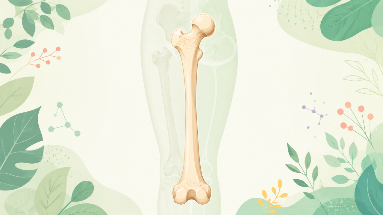
The femur has a long shaft that is cylindrical in shape with several curvatures along its length. Its proximal end, or head, is spheroidal and articulates with the acetabulum of the pelvis. At this end, you find two prominent bony protrusions known as the greater and lesser trochanters, which serve various muscle attachments.
The distal end consists of a condylar surface divided into medial (medial condyle) and lateral (lateral condyle) compartments. Each of these is covered by articular cartilage and articulates with the corresponding compartment of the tibia during knee flexion and extension.
The Function and Clinical Significance of the Femur
The primary function of the femur is to provide structural support, enable locomotion, and absorb shock during weight-bearing activities. In clinical contexts, injuries to the femur, such as fractures, can significantly impact a patient's mobility and quality of life. Understanding the anatomy and physiology of the femur is essential for medical professionals involved in diagnosis, treatment, and rehabilitation of such conditions.
The Femoral Neck and Greater Trochanter
Femoral Neck
The femoral neck is a narrow portion of the femur that connects the head to the shaft. It has an oblique orientation with the neck-shaft angle measuring approximately 125-130 degrees in adults. The femoral neck serves as the point of attachment for various muscles and ligaments, including the hip abductors and adductors.
Greater Trochanter
The greater trochanter is a large bony projection located on the lateral (outer) side of the proximal end of the femur. It plays an essential role in providing attachments for muscles involved in hip abduction, such as the gluteus medius and minimus. The greater trochanter can also be palpated through the skin when performing a physical examination.
The Femoral Shaft
The shaft of the femur is long and cylindrical, with various curvatures along its length. It serves as the weight-bearing portion of the bone during ambulation and provides attachment sites for muscles that act on the hip joint.
Medullary Cavity
Within the shaft of the femur lies the medullary cavity, which houses the marrow tissue. The medullary cavity is filled with red marrow in children and adolescents but gradually changes to yellow marrow as we age. This transformation occurs due to a shift in hematopoietic activity from red blood cell production to fat storage.
Muscle Attachments on the Femoral Shaft
The femoral shaft has multiple muscle attachment sites, including the:
- Iliacus (iliopsoas group)
- Pectineus
- Sartorius
- Adductor longus
- Gracilis
- Vastus lateralis, medialis, and intermedius
The Distal End of the Femur: Condyles and Trochlea
The distal end of the femur consists of two condyles (lateral and medial) separated by a trochlea. These structures facilitate knee flexion and extension.
Medial and Lateral Condyles
The medial and lateral condyles are rounded, articular surfaces on the distal end of the femur that articulate with the corresponding surfaces of the tibia during knee movement. The medial condyle is larger than the lateral one and has a more prominent anterior-posterior curvature.
Trochlea
The trochlea, or patellar surface, is a flat, longitudinal groove that runs along the medial border of the distal femur. It serves as the site of attachment for the patella during knee flexion and extension.
Conclusion
In conclusion, understanding the anatomy, morphology, physiology, and clinical significance of the femur is vital for those studying osteology. The femur plays a crucial role in weight-bearing, locomotion, and shock absorption, making it one of the most important bones in the human body.