Anatomy
course-show.h1-title
Discover the anatomy of the neck: from its bone and muscle structure to its major nerves and arteries. Learn to identify the different parts of the neck and understand their role in mobility, sensitivity, and protection of vital organs.
The neck, also known as the cervical region, is a vital part of the human body's anatomy that plays essential roles in supporting the head, facilitating movement, and housing important structures such as nerves and blood vessels. This comprehensive guide aims to provide an in-depth understanding of the neck's structure, functions, and clinical significance.
Introduction
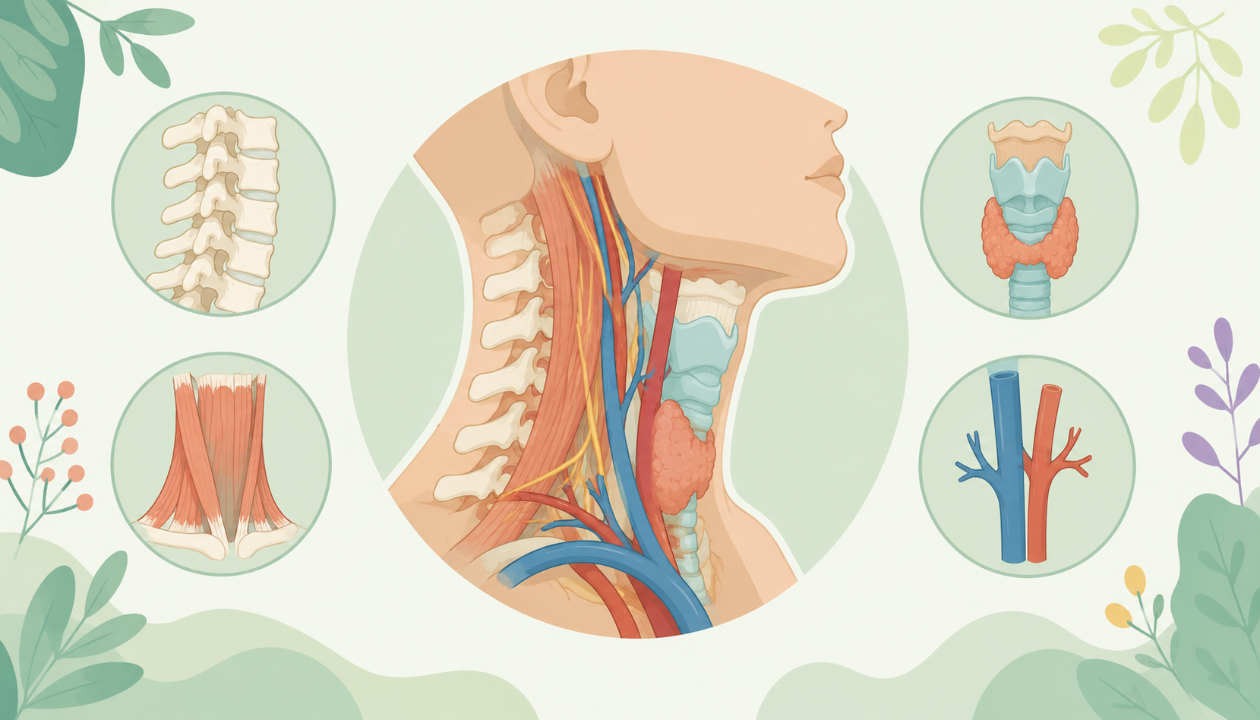
The neck, or cervical region, is the anterior portion of the vertebral column, extending from the base of the skull (occipital bone) to the thoracic inlet. It is divided into seven cervical vertebrae (C1-C7), each with unique morphological and functional characteristics. This introduction provides an overview of the neck's key features, importance, and organization for a more thorough exploration in subsequent sections.
Cervical Vertebrae
The seven cervical vertebrae are numbered from C1 to C7, with C1 (Atlas) and C2 (Axis) having specific roles in supporting the head and enabling rotation and flexion-extension movements. The remaining cervical vertebrae exhibit typical characteristics of spinal bones, such as a body, neural arch, pedicles, lamina, spinous process, transverse process, and facet joints.
Important Structures in the Neck
The neck houses essential structures like nerves, blood vessels, and muscles. The cervical spinal cord, an extension of the brainstem, runs through the vertebral canal and provides motor, sensory, and autonomic functions to various parts of the body. The cervical plexus, a network of nerves originating from the upper five or six cervical segments, also plays a crucial role in innervating structures such as the head, neck, diaphragm, and shoulder region.
In addition, numerous blood vessels travel through the neck, including the carotid arteries (common, internal, external), vertebral arteries, and jugular veins (internal and external). These vessels supply oxygenated blood to the head and brain while draining deoxygenated blood back to the heart.
Clinical Significance of the Neck
The neck's anatomy is crucial in understanding various clinical conditions, such as cervical spondylosis (osteoarthritis of the cervical vertebrae), herniated discs, and pinched nerves. In addition, injuries to the neck can potentially affect vital structures like the spinal cord or blood vessels, leading to symptoms such as pain, numbness, weakness, or even paralysis. Therefore, understanding the anatomy of the neck is essential for healthcare professionals involved in diagnosing, treating, and preventing these conditions.
Cervical Vertebrae in Detail
This section will delve deeper into each cervical vertebra (C1-C7), highlighting their unique characteristics and functions.
Atlas (C1)
The first cervical vertebra, or Atlas, is unique due to its lack of a body and the presence of two lateral masses connected by an anterior arch and a posterior arch. These masses articulate with the occipital condyles of the skull, allowing rotation and flexion-extension movements of the head.
Axis (C2)
The second cervical vertebra, or Axis, is also distinct due to its dens, a large, odontoid process projecting from its superior articular facet. The dens fits into the posterior arch of the Atlas (C1), creating the atlanto-axial joint, which facilitates rotation and allows for the forward flexion of the head.
Remaining Cervical Vertebrae (C3-C7)
The remaining cervical vertebrae (C3-C7) share several common features, including a body, neural arch, pedicles, lamina, spinous process, transverse process, and facet joints. However, they vary in size and shape, with C7 being the largest due to its role as the transition between the cervical and thoracic regions.
Important Structures of the Neck (Continued)
This section will explore additional essential structures found within the neck, such as muscles, ligaments, and the cervical plexus.
Muscles of the Neck
The muscles of the neck can be categorized into intrinsic and extrinsic muscles. Intrinsic muscles are located entirely within the neck and primarily provide mobility to the head. Extrinsic muscles originate outside the neck and insert into the skull, providing additional support and stability. Some examples include the sternocleidomastoid, scalene, and splenius muscles.
Ligaments of the Neck
Ligaments are dense connective tissues that connect bones to each other and provide stability to joints. In the neck, ligaments like the anterior atlanto-occipital membrane, transverse atlantal ligament, and alar ligaments help maintain the integrity of the atlanto-axial and atlanto-occipital joints.
Cervical Plexus
The cervical plexus is a network of nerves originating from the upper five or six cervical segments (C1-C5 or C6). It provides motor, sensory, and autonomic innervation to structures such as the head, neck, diaphragm, and shoulder region. Branches of the cervical plexus include the greater occipital nerve, phrenic nerve, ansa cervicularis, and the cervical branches that supply sensation to the skin and muscles of the neck and shoulder area.
Clinical Conditions Affecting the Neck
This section will discuss various clinical conditions affecting the neck, such as cervical spondylosis, herniated discs, and pinched nerves.
Cervical Spondylosis
Cervical spondylosis is a common degenerative condition affecting the intervertebral discs and facet joints of the cervical vertebrae. It can cause neck pain, stiffness, and radiating symptoms such as numbness or weakness in the arms.
Herniated Disc (Cervical Radiculopathy)
A herniated disc occurs when the inner gel-like substance of an intervertebral disc bulges out through a tear in its outer layer, potentially compressing nearby nerves and causing symptoms such as pain, numbness, or weakness in the affected arm.
Pinched Nerve (Cervical Foraminal Stenosis)
Pinched nerve, also known as cervical foraminal stenosis, occurs when a nerve root is compressed as it exits the foramen (neural opening) between two adjacent cervical vertebrae. This can cause symptoms such as neck pain, arm pain, numbness, or weakness in the affected arm.
Conclusion
Understanding the anatomy and functions of the neck is essential for healthcare professionals involved in diagnosing, treating, and preventing various clinical conditions affecting this region. This comprehensive guide has provided a detailed overview of the cervical vertebrae, important structures within the neck, and common clinical conditions affecting this vital area.