Cellular Biology
The peroxisomes
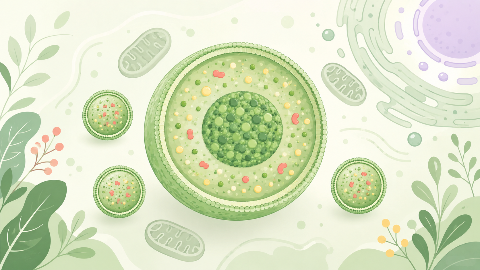
Discover peroxisomes, small cellular organelles that are key to the survival and adaptation of our cells! In this cell biology course, you'll explore their structure...
Arthrology, or syndesmology
Delving into the study of human joints, this course will introduce you to the hip joint, a key component of the musculoskeletal system. We'll explore the structures that make it up and the movements it enables. We'll also develop our understanding of the pathologies that can affect this crucial joint.
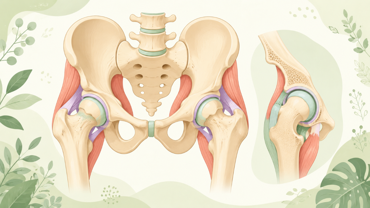
The hip joint, or coxal-femoral joint, is one of the largest and most robust synovial joints in the human body. It serves as a ball-and-socket joint connecting the pelvis (acetabulum) to the femur (head). The hip joint enables a wide range of movements, including flexion, extension, abduction, adduction, internal rotation, and external rotation. This article provides an in-depth exploration of the anatomy, development, biomechanics, pathologies, and clinical considerations related to the hip joint.
The bony elements forming the hip joint include the acetabulum (part of the pelvis) and the femoral head. The acetabulum is a deep cup-shaped cavity formed by the ilium, ischium, and pubis. The femoral head is the rounded end of the femur located above the neck.
The articular surface of the acetabulum is lined with hyaline cartilage that covers the bony cavity's labrum. The articular surface of the femoral head is also covered by hyaline cartilage, which articulates with the acetabular labrum and socket.
Several ligaments provide stability to the hip joint, including:
The hip joint develops from three embryonic tissues: the somatic mesoderm, sclerotome, and lateral plate mesoderm. By the end of week 5, the lower limb bud appears as an outgrowth from the caudal end of the body. The femur and pelvis differentiate and form the hip joint during the following weeks.
The hip joint's range of motion is essential for locomotion, posture maintenance, and other daily activities. The primary muscles responsible for hip movements are:
Injuries to the hip joint can result from falls, sports-related accidents, or high-impact activities. Common injuries include:
Osteoarthritis (OA) is the most common joint disorder, characterized by degeneration of articular cartilage, subchondral bone changes, and osteophyte formation. Risk factors for developing hip OA include aging, obesity, genetics, and previous joint injuries.
DDH is a congenital or developmental disorder characterized by an unstable hip joint due to abnormal acetabular development. Early detection and treatment are crucial to prevent further complications such as limb length discrepancy, osteoarthritis, and reduced mobility.
Proper diagnosis and management of hip-related disorders require a thorough understanding of the hip joint's anatomy, function, and potential pathologies. A combination of clinical examination, imaging modalities (X-ray, MRI), and arthroscopy can help in making an accurate diagnosis. Treatment options may include:
Penses-tu tout connaître de ce cours ? Ne tombe pas dans les pièges, entraine-toi à l'aide des QCM ! eBiologie recense des centaines de questions pour t'aider à maîtriser ce sujet.
Discover peroxisomes, small cellular organelles that are key to the survival and adaptation of our cells! In this cell biology course, you'll explore their structure...
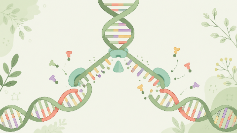
Discover how our DNA replicates with each cell division in this molecular biochemistry course: "DNA Replication." You'll learn the key steps in this crucial process...
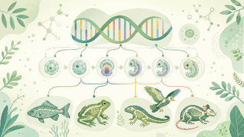
Learn about evolutionary developmental biology, the field that studies the mechanisms of embryonic development and their evolution at the molecular, cellular, and st...