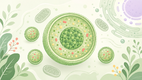
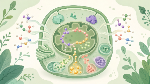
Os peroxissomos
Descubra os peroxsomos, pequenos organelos celulares que são fundamentais para a sobrevivência e adaptação de nossas células! Neste curso de biologia celular, você e...
Histology
Discover the anatomy and function of striated muscle tissue in this histology course! This subject explores the different cellular structures, muscle contraction, and coordination between these muscles. Deepen your knowledge of the role of these tissues in body mobility as well as their ultrastructural structure at the microscopic level.

The striated muscles, also known as skeletal muscles, are the type of muscle that is attached to bones and is responsible for body movements. In this course, we will delve into the anatomy, histology, physiology, and function of these essential tissues in vertebrates.
Striated muscles are found throughout the bodies of vertebrates. They are named striated because they have a distinct banding pattern when viewed under a microscope, resulting from the arrangement of contractile proteins.
There are two main types of striated muscles: skeletal (also known as voluntary) and cardiac (involuntary). Skeletal muscles are attached to bones through tendons and are responsible for movement, while cardiac muscles are found in the heart and contract rhythmically to pump blood.
A muscle fiber is composed of long, cylindrical cells with centrally located nuclei. The cell membrane (sarcolemma) surrounds each muscle fiber. Inside the muscle fiber are numerous myofibrils that run parallel to one another and contain sarcomeres—the functional units of striated muscles.
The sarcomere is further divided into two regions: the A-band (anisotropic band) and the I-band (isotropic band). The A-band consists of thick filaments (myosin) and overlapping thin filaments (actin), while the I-band contains only thin filaments. The H-zone, found within the A-band, is devoid of thin filaments.
The striped appearance of striated muscles results from the alternating light and dark bands observed in sarcomeres under a microscope. The light A-bands represent the myosin thick filaments, while the dark I-bands contain only actin thin filaments. The thin, light-colored Z-lines run parallel to each other along the length of the sarcomere and provide structural support.
The process of muscle contraction involves the interaction between myosin and actin filaments. During relaxation, myosin heads bind ATP, while in contraction, they release ATP and bind to actin filaments, causing them to slide along the thick filaments, leading to shortening of the sarcomere and overall muscle fiber.
The degree of muscle contraction is regulated by the interaction between calcium ions (Ca²⁺) and troponin-tropomyosin complexes in the thin filaments. In the presence of Ca²⁺, the tropomyosin molecule shifts position, exposing the myosin-binding sites on actin, allowing for muscle contraction to occur.
Striated muscles are crucial for movement and maintaining posture by contracting in response to nerve signals. They generate force and produce movements in various directions, allowing for complex actions like walking, grasping objects, and speaking.
The force generated by a muscle is proportional to the number of active muscle fibers and the degree of their contraction. Muscle fatigue occurs when muscles are exercised beyond their capacity, leading to decreased force generation and eventual failure.
Understanding the structure, function, and regulation of striated muscle tissue provides valuable insights into the intricate workings of the muscular system and its role in overall body function. Further research in this area may lead to improved treatments for muscle-related disorders and enhance our understanding of physical performance and exercise physiology.
Do you think you know everything about this course? Don't fall into the traps, train with quizzes! eBiologie has hundreds of questions to help you master this subject.


Descubra os peroxsomos, pequenos organelos celulares que são fundamentais para a sobrevivência e adaptação de nossas células! Neste curso de biologia celular, você e...
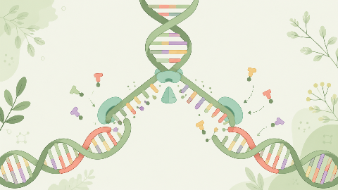
Descubra como nosso DNA se replica a cada divisão celular neste curso de bioquímica molecular: "Replicação do DNA." Você aprenderá os passos principais desse process...
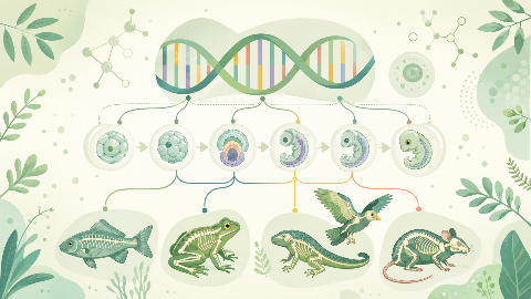
Aprenda sobre biologia evolutiva do desenvolvimento, o campo que estuda os mecanismos do desenvolvimento embrionário e sua evolução nos níveis molecular, celular e e...
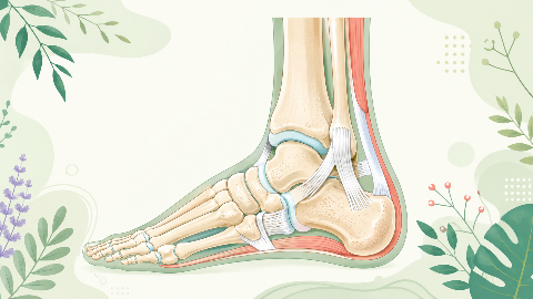
Descubra a articulação talonípede neste curso de sindemologia: os ossos, cartilagem e ligamentos que possibilitam a articulação dessa articulação complexa. Aprenda a...
Descubra o metabolismo dos aminoácidos neste curso empolgante e técnico! Você vai aprender a identificar os diferentes tipos de aminoácidos e entender seu papel esse...
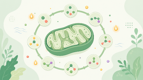
Descubra o ciclo de Krebs: o processo metabólico central que converte ácidos graxos em energia para a célula. Entenda as etapas desse processo e aprenda como ele int...
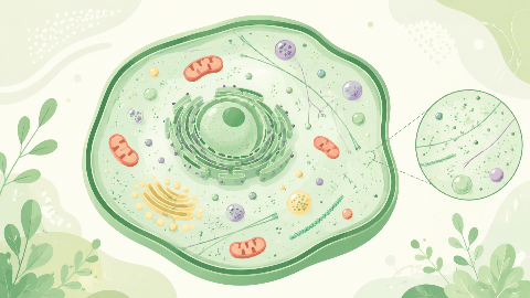
Mergulhe no intrigante mundo dos Compartimentos Celulares! Explore a estrutura, função e dinâmica do citosol, incluindo seu papel na síntese de proteínas, produção d...
