Myology
course-show.h1-title
In this myology course, we explore the calf muscles, which play a vital role in our body's mobility and stability. We study their anatomy, function, and control, as well as their interactions with other muscle structures. With this in-depth knowledge of the calf muscles, we can better understand the origins and treatments of calf-related disorders.
Introduction
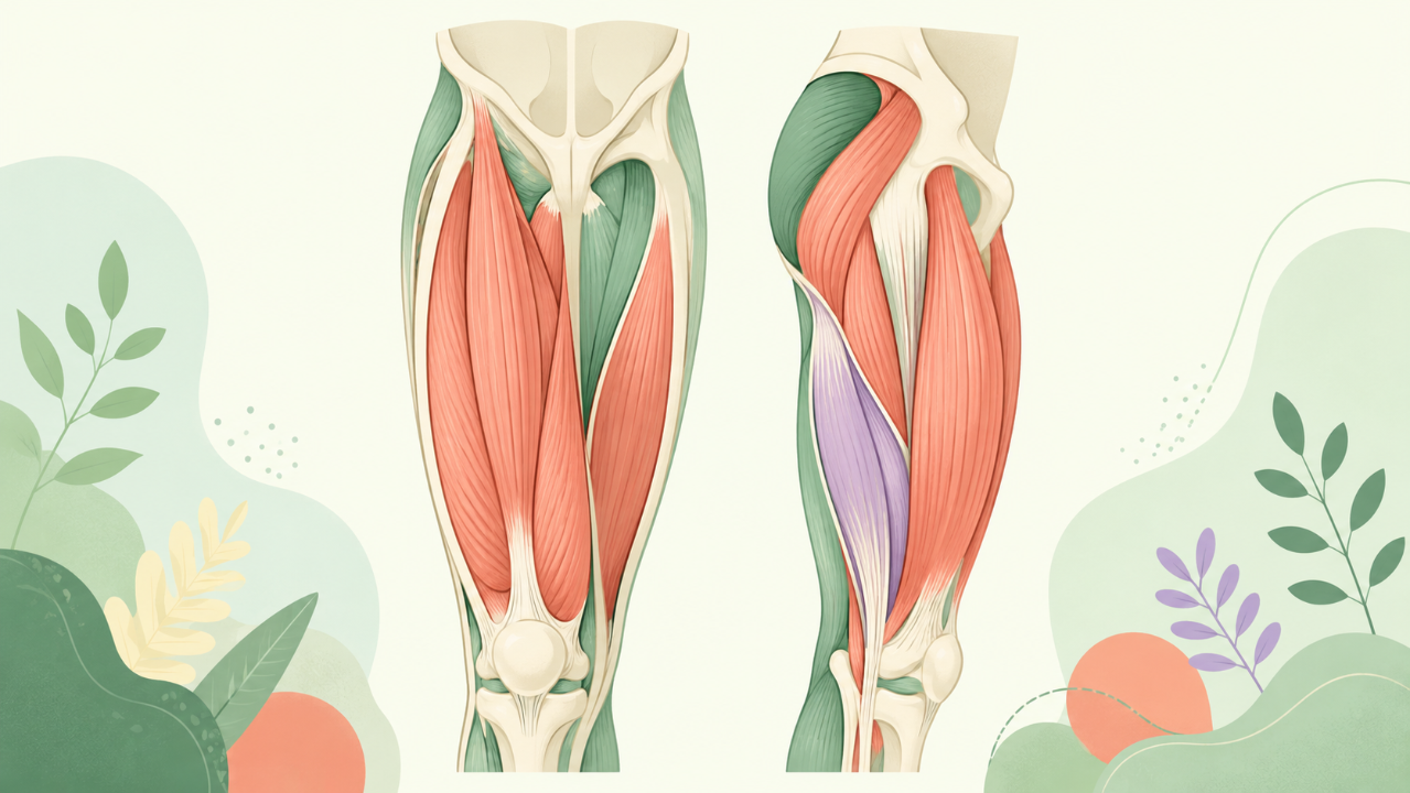
The human thigh is a vital region of the musculoskeletal system, housing several essential muscle groups that enable locomotion, stability, and postural control. This comprehensive course delves into an in-depth analysis of the muscles constituting the thigh, highlighting their functions, origins, insertions, actions, innervations, and relationships with other structures.
Anatomical Overview
Regions of the Thigh
The human thigh is divided into three regions: anterior, posterior, and medial. Each region houses unique muscle groups responsible for different movements and functions.
- Anterior Region: Home to quadriceps femoris, sartorius, and pectineus muscles.
- Posterior Region: Contains the hamstrings (semitendinosus, semimembranosus, and biceps femoris) and popliteus muscles.
- Medial Region: Accommodates adductors - a group of six muscles responsible for thigh adduction.
Quadriceps Femoris
Function & Anatomy
The quadriceps femoris muscle group is primarily responsible for knee extension and stabilization during various movements, such as walking, running, and jumping. It consists of four muscles: rectus femoris, vastus lateralis, vastus intermedius, and vastus medialis. Each has distinct origins, insertions, and actions within the leg joints.
Origin & Insertion
- Rectus Femoris: Originates from the anterior ilium and the pubis (iliac fossa), and inserts on the patella and quadriceps tendon, which attaches to the tibial tuberosity.
- Vastus Lateralis: Arises entirely from the femur's lateral intermuscular septum and the greater trochanter, and inserts onto the patella and quadriceps tendon.
- Vastus Intermedius: Originates from the intermuscular septum between the vastus lateralis and rectus femoris and the lesser trochanter of the femur, inserting into the same tendons as the other two muscles.
- Vastus Medialis: Originates from the medial intermuscular septum and the adductor magnus, and inserts onto the same tendons as the other quadriceps muscles.
Hamstrings
Function & Anatomy
The hamstrings consist of three muscles - semitendinosus, semimembranosus, and biceps femoris. They primarily function in knee flexion (bending) and hip extension (straightening). Each muscle has distinct origins, insertions, and actions within the leg joints.
Origin & Insertion
- Semitendinosus: Originates from the ischial tuberosity and the medial part of the linea aspera on the femur, inserting onto the tibial collateral ligament at the medial aspect of the knee joint.
- Semimembranosus: Arises from the ischial tuberosity and the posterior surface of the femur (linea aspera), and inserts onto the posterior surface of the medial tibia and the head of the fibula.
- Biceps Femoris: Consists of a long head and a short head. The long head originates from the ischial tuberosity, while the short head arises from the femur's lateral intermuscular septum. Both heads converge to form a common tendon, which inserts onto the head of the fibula and the bicipital aponeurosis at the lateral aspect of the knee joint.
Popliteus
Function & Anatomy
The popliteus muscle is essential for rotating the tibia during flexion, stabilizing the knee joint, and facilitating the transition from heel strike to mid-stance in locomotion. It originates from the lateral femoral condyle and inserts on the fibular head.
Adductors
Function & Anatomy
The adductor group is responsible for thigh adduction, medial rotation of the leg, and maintaining the pelvis' stability during various movements. They consist of six muscles - gracilis, pectineus, adductor longus, adductor brevis, adductor magnus (long head), and adductor magnus (short head).
Origin & Insertion
- Gracilis: Originates from the anterior 2/3 of the ramus of the pubis and inserts onto the linea aspera on the medial aspect of the proximal tibia.
- Pectineus: Arises from the pecten pubis, inferior pubic ramus, and obturator membrane, and inserts onto the adductor longus tendon and the linea aspera on the femur's medial condyle.
- Adductor Longus: Originates from the pubic ramus and the ramus of the ischium, inserting onto the linea aspera on the medial aspect of the proximal tibia.
- Adductor Brevis: Originates from the ramus of the pubis, the ischium, and the obturator membrane, and inserts onto the superior surface of the linea aspera on the femur's medial condyle.
- Adductor Magnus (Long Head): Arises from the ischial tuberosity and the adductor magnus brevis, inserting onto the linea aspera on the posterior aspect of the femur's lateral condyle.
- Adductor Magnus (Short Head): Originates from the ischial tuberosity and inserts onto the medial aspect of the proximal fibula.
Innervations & Relationships with Other Structures
Innervation
- Quadriceps Femoris: Primarily innervated by the femoral nerve (L2-L4).
- Hamstrings: Primarily innervated by the sciatic nerve, which is a combination of the tibial (S1-S3) and common peroneal (L4-S2) nerves.
- Popliteus: Innervated by the tibial nerve (L4-L6).
- Adductors: Primarily innervated by the obturator nerve (L2-L4) and the anterior division of the femoral nerve (L2-L4).
Relationships with Other Structures
Thigh muscles share close relationships with various structures within the leg joints, tendons, ligaments, and aponeuroses. Understanding these relationships is crucial for understanding their functions, movements, and potential injuries.
Clinical Significance
Understanding the anatomy, function, and relationships of thigh muscles is essential in diagnosing and treating related injuries, as well as developing effective exercise programs for rehabilitation or athletic performance enhancement. Common injuries affecting these muscles include strains, sprains, contusions, tendinitis, and tears.
Conclusion
This comprehensive course has explored the muscles of the thigh, highlighting their unique functions, origins, insertions, actions, innervations, and relationships with other structures. By gaining a deeper understanding of these essential muscle groups, students can better appreciate the complexity of human anatomy, the intricacies of the musculoskeletal system, and how to apply this knowledge in clinical practice or exercise programming.