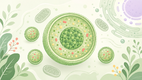
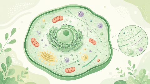
The peroxisomes
Discover peroxisomes, small cellular organelles that are key to the survival and adaptation of our cells! In this cell biology course, you'll explore their structure...
Osteology
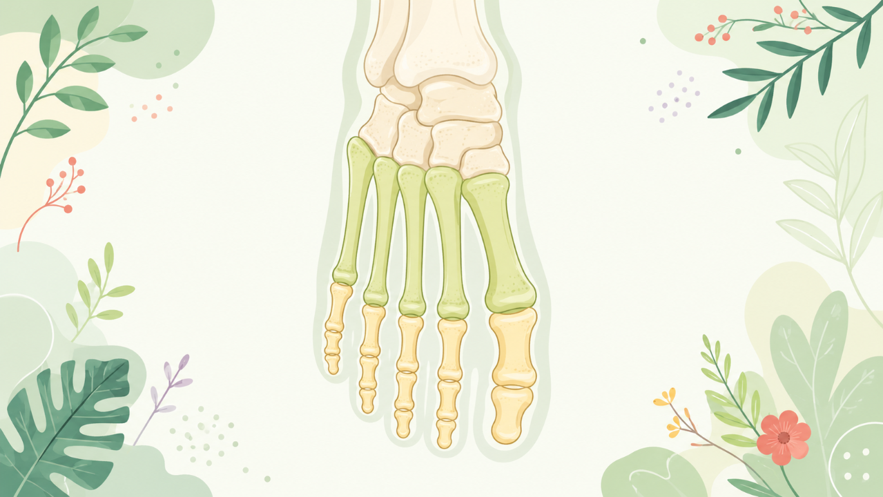
Discover the metatarsal and phalangeal bones of your foot: their structures, functions, and importance for human locomotion. This osteology course will help you understand the role of these bones in our skeletal system, while exploring their evolution and the deformities that can occur.
This comprehensive academic course aims to provide an in-depth exploration of the metatarsal and phalange bones, key components of the human foot anatomy. This study falls under the category of Osteology, the branch of anatomy that deals with the skeletal system. The objective is to equip students with a thorough understanding of these structures, their functions, development, and clinical relevance.
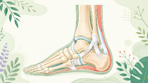
Before diving deep into the metatarsals and phalanges, it is essential to understand the overall structure of the foot skeleton. The human foot consists of three sections: the hindfoot, midfoot, and forefoot. The metatarsals and phalanges belong to the forefoot region.
The hindfoot contains the talus (talus bone), calcaneus (heel bone), and navicular and cuboid bones of the midfoot. These bones provide stability during weight-bearing activities, serving as a connection point between the leg and the foot.
The forefoot consists of five long, slender bones called metatarsals. Numbered from medial to lateral, they are labeled as the first through fifth metatarsals. These bones extend from the tarsometatarsal joint in the midfoot to the base of the toes at the metatarsophalangeal (MTP) joint. Each metatarsal bone articulates with both a proximal tarsal bone and a distal phalanx.
The metatarsals have an elongated, cylindrical shape with a slightly expanded head region at their proximal ends. The shaft is long and slender, while the distal end tapers to a smaller, rounded surface that forms the base for the phalanges. The metatarsals play a crucial role in weight-bearing activities, absorbing impact forces during locomotion and helping to maintain balance and propel the body forward.
Each digit (toe) in the human foot contains three phalanges: a proximal, intermediate, and distal phalanx. These bones are small, slender, and tapered in shape, with smooth, rounded articular surfaces for joint connections. The phalanges articulate with one another to form interphalangeal (IP) joints and the metatarsophalangeal (MTP) joint with the respective metatarsal bone.
The proximal phalanx is the largest and most robust of the three, providing a stable base for the toe. It connects to the metatarsal at the MTP joint and bears weight during walking and standing. The intermediate phalanx is shorter and thinner than the proximal phalanx and plays a supporting role in maintaining toe stability. The distal phalanx is the smallest bone of the toe, responsible for tipping the toe upward when needed, such as during pushing off during walking or running.
The development of the metatarsals and phalanges begins in the embryonic stage, when the limbs differentiate into distinct regions: the humerus region (upper arm), femur region (thigh), forelimb (hand/arm) and hindlimb (leg/foot). The metatarsals and phalanges are derived from the limb bud, which eventually forms the structures of the hand and foot.
During this phase, the limb bud undergoes a process called regionalization, where specific regions along the length of the bud develop distinct characteristics. The forelimb region (zone of polarizing activity) generates the metatarsals and phalanges.
Subsequent to regionalization, mesenchymal cells (precursor cells) in the limb bud begin to differentiate into chondroblasts, which form cartilage templates for future bone development. By weeks 6 to 8, the metatarsals and phalanges are evident as small cartilaginous structures.
Ossification begins in the center of each long bone (primary ossification center) during the ninth week of embryonic development. The primary centers for the metatarsals and phalanges are formed at their diaphyseal regions, eventually growing towards the ends of the bones through a process called endochondral ossification.
Injuries or disorders affecting the metatarsals and phalanges can significantly impact an individual's ability to walk, run, or perform other activities requiring the use of their feet. Common conditions that affect these bones include fractures, dislocations, osteoarthritis, hammertoe, and claw toe. Understanding the normal anatomy and function of these structures helps medical professionals diagnose and treat such conditions effectively.
This course has provided a comprehensive exploration of the metatarsal and phalange bones in the human foot. By understanding their structure, function, development, and clinical relevance, students can develop a strong foundation for further studies in osteology and related fields.
Do you think you know everything about this course? Don't fall into the traps, train with quizzes! eBiologie has hundreds of questions to help you master this subject.
Discover peroxisomes, small cellular organelles that are key to the survival and adaptation of our cells! In this cell biology course, you'll explore their structure...
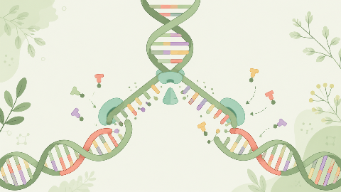
Discover how our DNA replicates with each cell division in this molecular biochemistry course: "DNA Replication." You'll learn the key steps in this crucial process...
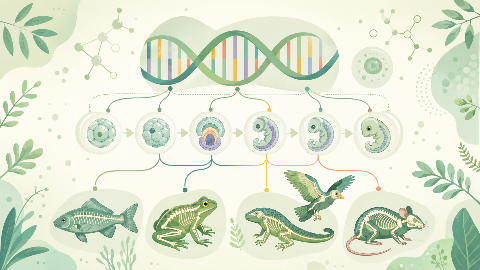
Learn about evolutionary developmental biology, the field that studies the mechanisms of embryonic development and their evolution at the molecular, cellular, and st...
Discover the talonipedal joint in this syndesmology course: the bones, cartilage, and ligaments that enable the articulation of this complex joint. Learn to identify...
Discover amino acid metabolism in this exciting and technical course! You'll learn to identify the different types of amino acids and understand their essential role...
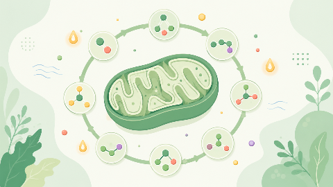
Discover the Krebs cycle: the central metabolic process that converts fatty acids into energy for the cell. Understand the steps in this process and learn how it int...
Dive into the intriguing world of Cellular Compartments! Explore the cytosol's structure, function, and dynamics, including its role in protein synthesis, energy pro...
