Embryology or developmental biology.
course-show.h1-title
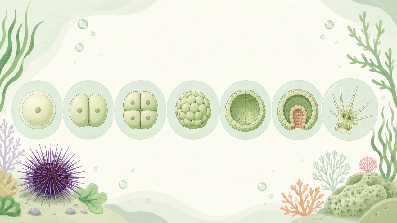
Discover the evolution of starfish embryos: a keen view of embryonic development! Explore the key stages of the process, including cleavage, gastrulation, and morphogenesis. Also enjoy in-depth studies of the key genes involved in this spectacular natural phenomenon.
Introduction
Sea urchins, a group of marine echinoderms, are valuable models for studying embryonic development due to their simple yet complex organization and transparent eggs. This course will delve into the intricacies of embryonic development in sea urchins, providing an in-depth understanding of this fascinating process.
Overview
The sea urchin life cycle consists of two main stages: the diploid adult stage and the haploid planula larva stage. Embryonic development is the transition from fertilized egg to planula, a free-swimming larva that eventually metamorphoses into an adult sea urchin. This process involves numerous cellular and molecular events that will be explored in detail.
Importance of Sea Urchins as Model Organisms
Sea urchins have been extensively used in developmental biology research for over a century due to their numerous advantages, including:
- Transparent eggs: The transparent eggs allow direct visualization of the developing embryo without the need for microscopy.
- Simplicity: Sea urchin embryos have fewer cells than many other model organisms, making it easier to study cellular processes and their coordination.
- Conserved mechanisms: Many fundamental developmental processes in sea urchins are conserved across metazoan phyla, making discoveries relevant for understanding human development as well.
- Accessibility: Sea urchins are abundant in many coastal regions and can be easily collected for research purposes.
Fertilization and Cleavage
Fertilization
Fertilization occurs when a sperm cell penetrates the egg, triggering a series of events that lead to embryonic development. In sea urchins, fertilization results in the activation of the maternal genome and the initiation of transcription.
Cleavage
Cleavage is the rapid succession of mitotic cell divisions that follow fertilization. Sea urchin eggs undergo a series of cleavages, resulting in a blastula stage with thousands of cells arranged in multiple layers. Each cleavage division follows a specific pattern, either radial or spiral.
Gastrulation
Blastula to Gastrula Transformation
Gastrulation is the process by which the blastula transforms into a gastrula, forming three primary germ layers: endoderm, mesoderm, and ectoderm. This transformation occurs through the movements of cells, known as invagination and ingression.
Invagination and Ingression
Invagination is the inward movement of cells from the animal hemisphere, forming the archenteron or blastopore. Ingression is the movement of cells into the archenteron, forming the primitive streak and giving rise to the mesoderm and endoderm germ layers.
Axis Formation
The embryo acquires an anterior-posterior (AP) axis during gastrulation due to the formation of the primary axes: the animal-vegetal (AV) axis and the dorsal-ventral (DV) axis. These axes provide positional information for the development of specific tissues and organs.
Animal-Vegetal Axis
The AV axis forms during fertilization, with the sperm entering from the animal pole (AP) and the vegetal pole (VP) located at the opposite end of the egg. The first cell divisions result in a mosaic of cells that retain their position along the AV axis.
Dorsal-Ventral Axis
The formation of the DV axis occurs during gastrulation, with the dorsal side (D) being the future site of the archenteron and the ventral side (V) being the eventual ectoderm. The establishment of this axis is regulated by the positioning of pigment granules in the egg and the migration of cells during invagination and ingression.
Organogenesis
Organogenesis is the process by which the three germ layers give rise to various organs and tissues throughout the embryo. This stage follows gastrulation and continues until metamorphosis into the adult sea urchin.
Ectoderm
The ectoderm gives rise to several structures, including the epidermis, nerve tissue, and sensory organs. The ectoderm undergoes a series of morphogenetic movements, such as epithelial-mesenchymal transition (EMT), to form these tissues.
Mesoderm
The mesoderm forms the musculature, skeletal structures, and circulatory system of the sea urchin. The mesoderm undergoes a process called convergent extension, where cells migrate towards the midline and elongate to form these tissues.
Endoderm
The endoderm gives rise to the digestive tract and other internal organs, such as the gonads. The development of the endoderm involves the coordination of cell movements and signaling events between neighboring cells.
Conclusion
This course has provided an overview of embryonic development in sea urchins, covering fertilization, cleavage, gastrulation, axis formation, organogenesis, and more. The simplicity and accessibility of these model organisms make them ideal for studying the fundamental mechanisms of embryonic development.