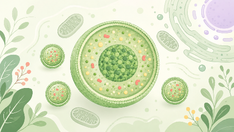
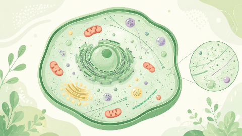
The peroxisomes
Discover peroxisomes, small cellular organelles that are key to the survival and adaptation of our cells! In this cell biology course, you'll explore their structure...
Myology
Discover the function, anatomy, and physiology of abdominal muscles in this myology course. You'll explore the various types of muscle fibers present, their role in postural stability, and the coordinated actions they perform during body movements.
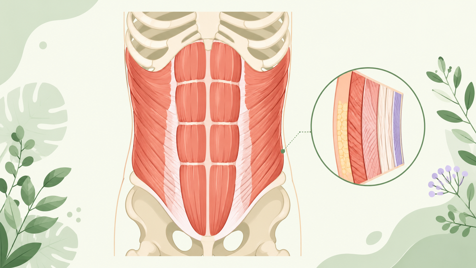
This comprehensive academic course provides an in-depth exploration of the abdominal muscles, a vital component of the human musculoskeletal system within the field of Myology. The focus is on presenting a clear, detailed, and rigorous understanding of the structure, function, and significance of these muscles in human physiology.
The abdomen is the part of the body between the thorax and pelvis. It is bounded by the following:
The abdominal muscles can be divided into three groups:
Each group will be discussed in detail, including their origins, insertions, actions, and innervation.
The external abdominal muscles consist of four layers:
The internal abdominal muscles include three layers:
Understanding the anatomy and function of the abdominal muscles is crucial for healthcare professionals, physical therapists, personal trainers, and athletes. Common conditions related to these muscles include hernias, strains, and sports injuries.
This course has provided a comprehensive exploration of the abdominal muscles, delving into their anatomy, function, innervation, and clinical relevance within the field of Myology.
Do you think you know everything about this course? Don't fall into the traps, train with quizzes! eBiologie has hundreds of questions to help you master this subject.
Discover peroxisomes, small cellular organelles that are key to the survival and adaptation of our cells! In this cell biology course, you'll explore their structure...
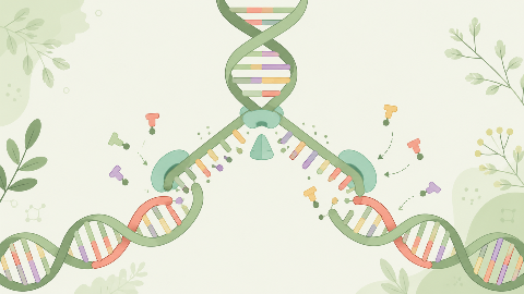
Discover how our DNA replicates with each cell division in this molecular biochemistry course: "DNA Replication." You'll learn the key steps in this crucial process...
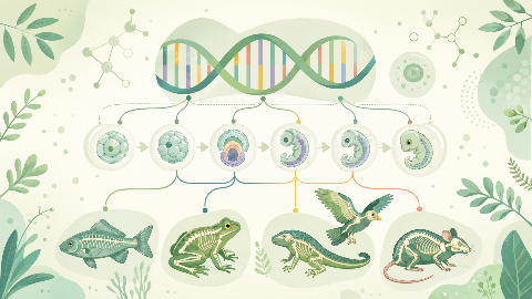
Learn about evolutionary developmental biology, the field that studies the mechanisms of embryonic development and their evolution at the molecular, cellular, and st...
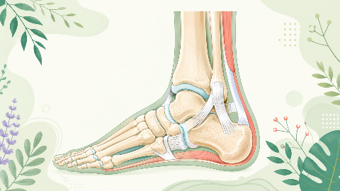
Discover the talonipedal joint in this syndesmology course: the bones, cartilage, and ligaments that enable the articulation of this complex joint. Learn to identify...
Discover amino acid metabolism in this exciting and technical course! You'll learn to identify the different types of amino acids and understand their essential role...
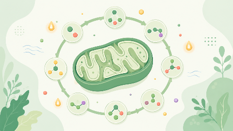
Discover the Krebs cycle: the central metabolic process that converts fatty acids into energy for the cell. Understand the steps in this process and learn how it int...
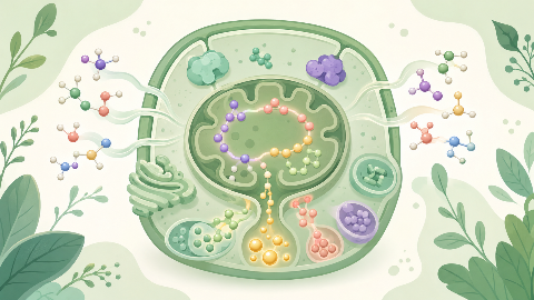
Dive into the intriguing world of Cellular Compartments! Explore the cytosol's structure, function, and dynamics, including its role in protein synthesis, energy pro...
