Osteology
course-show.h1-title
Explore the intricate world of bone biology in our engaging Osteology course! Delve into the structure and function of bones, the cellular makeup of bone tissue, and the developmental processes that shape our skeletal system.
Introduction
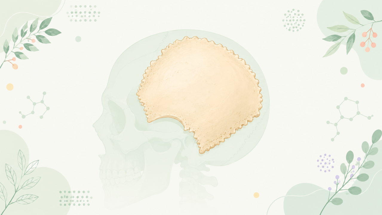
This comprehensive course is designed to provide advanced undergraduate students with an extensive and systematic understanding of the parietal bone, a significant cranial bone in vertebrate skeletons. The focus will be on its structure, function, development, and clinical relevance within the broader context of osteology.
Background Information
Definition and Classification
The parietal bone is one of the eight bones that constitute the cranium or skull, specifically part of the neurocranium. It can be found on either side of the cranial cavity, forming the lateral walls and roof.
Importance and Significance
Understanding the parietal bone is crucial for several reasons:
- It plays a vital role in protecting the brain from external trauma and internal pressure.
- The parietal bone's morphology and variations are useful in taxonomic studies, particularly in forensic anthropology and evolutionary biology.
- Clinical significance lies in its involvement in various skull fractures and deformities, as well as its role in craniometrics for determining age and sex.
Anatomy and Morphology of the Parietal Bone
Gross Anatomy
The parietal bone can be divided into three main parts: the squamosal, frontal, and temporal parts. These regions are connected by sutures, allowing for growth during development.
- The squamosal part forms the posterior part of the temporal bone and articulates with the petrosal part of the temporal bone, the mastoid process, and the occipital bone.
- The frontal part meets the frontal bone at the coronal suture and contributes to the lateral wall of the orbital cavity.
- The temporal part forms the greater part of the side walls of the cranial cavity and is involved in articulation with several other bones, including the sphenoid, zygomatic, maxilla, and palatine bones.
Microscopic Anatomy
The parietal bone consists primarily of compact bone, which is dense, hard, and forms the outer layer of most bones. The internal structure contains numerous small canals for blood vessels and nerves known as haversian systems or osteons.
Development and Ossification of the Parietal Bone
Embryology and Ontogeny
The parietal bone originates from the paraxial mesoderm, specifically the medial and lateral somites. Its development can be divided into two stages: primary ossification centers and secondary ossification centers.
- The primary ossification center appears around the fourth week of gestation, located at the junction of the squamosal and frontal parts of the parietal bone.
- Secondary ossification centers form later, around the seventh week, in the temporal part of the parietal bone. These regions will fuse with the primary center during development.
Ossification Centers' Fusion and Completion
Fusion between ossification centers takes place gradually over time:
- The squamosal and frontal parts of the parietal bone fuse by the end of the first year, resulting in a single, continuous plate-like structure on each side.
- The temporal part will subsequently fuse with the rest of the parietal bone during late childhood or early adolescence.
Clinical Relevance and Pathologies
Skull Fractures and Deformities
The parietal bone is commonly involved in skull fractures, particularly linear and diastatic fractures, which can have varying degrees of severity depending on the location, extent, and cause.
- Linear fractures are transverse breaks along the sutures or the outer table of the bone, often resulting from mild trauma.
- Diastatic fractures involve disruption of the sutures between bones, primarily affecting young individuals whose sutures have not yet fused completely.
Craniometrics and Forensic Anthropology
The parietal bone plays an essential role in craniometrics, a technique used to estimate age, sex, and ancestry from skull measurements. The shape, thickness, and texture of the parietal bone can provide valuable insights into an individual's biological profile.
Conclusion
Understanding the parietal bone is pivotal for comprehending the intricate structure and function of vertebrate crania. This course has offered a detailed exploration of its morphology, development, clinical relevance, and significance in various scientific fields.