Osteology
course-show.h1-title
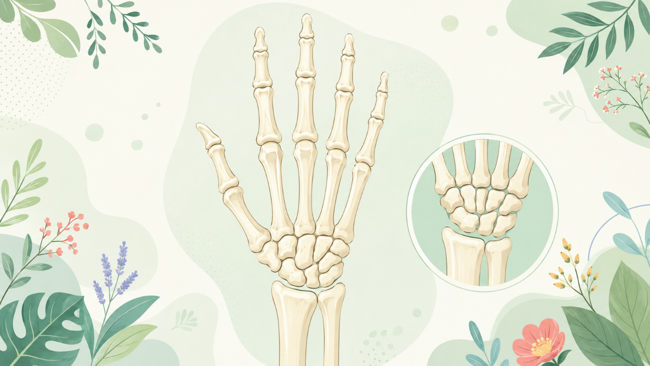
Discover the osteology of the wrist in this comprehensive course! An in-depth study of the bones of the hand will teach you how to identify key bone structures and understand their functional roles.
Introduction
The study of bones is essential in understanding the function, evolution, and diversity of various organisms. This course focuses on the osteology of the human hand, a complex and intricate part of the upper limb that plays a crucial role in our daily activities. The hands, composed of 27 bones arranged in a specific pattern, are remarkable examples of bone adaptation to function.
An Overview of Hand Bones
The Carpals
The carpal bones, located at the wrist, comprise eight bones organized into two rows. They include:
- Scaphoid
- Lunate
- Triquetral
- Pisiform
- Trapezium
- Trapezoid
- Capitate
- Hamate
The Metacarpals
The metacarpal bones are five long, flat bones that connect the carpals to the phalanges (finger bones). They are numbered from I to V, corresponding to the thumb to little finger.
The Phalanges
There are 14 phalanges in total, with each digit (excluding the thumb) having three phalanges: a proximal, middle, and distal phalanx. The thumb has only two phalanges due to its unique structure and function.
Bone Structure and Function
Diaphysis, Epiphysis, and Metaphysis
Each long bone consists of three regions: diaphysis, epiphysis, and metaphysis. Understanding these regions is essential for comprehending the growth, remodeling, and fracture patterns of hand bones.
- Diaphysis: The shaft or central portion of a long bone. This region contains compact bone and performs weight-bearing functions.
- Epiphysis: The ends of long bones are referred to as epiphyses. These regions consist of spongy bone, cartilage, and growth plates (epiphyseal plates). They allow for bone elongation during development.
- Metaphysis: Transitional zones between the diaphysis and epiphyses, containing both compact and spongy bone.
Articulations of Hand Bones
The hand bones are connected by various types of joints that enable flexibility, stability, and function. The most common joints found in the hand are:
- Synovial joints: These diarthroses allow for a wide range of motion through the articulation of articular cartilage-covered surfaces. Examples include interphalangeal, metacarpophalangeal, and carpometacarpal joints.
- Condylar joints: These joints, found between the distal end of the radius and the proximal row of carpal bones (radiocarpal joint), enable slight mobility while providing stability to the wrist.
- Sesamoid joints: Small articulations found within tendons, such as those between the first metacarpal and the trapezium and trapezoid in the thumb, facilitate smooth movement during action.
Developmental Aspects of Hand Bones
Hand bone development is a complex process influenced by genetic factors, intrauterine positioning, and postnatal environmental conditions. Understanding this development is essential for diagnosing various congenital anomalies and developmental disorders affecting the hand.
- Stages of development: The development of the hand bones can be divided into four main stages:
- Embryonic stage: During weeks 3 to 5, the limb buds differentiate into the upper and lower limbs.
- Formative stage: From weeks 6 to 12, the limbs further develop, with the formation of digits, carpal bones, and metacarpal bones.
- Pre-ossification stage: After 12 weeks, the cartilaginous models of hand bones ossify, starting with the phalanges and ending with the metacarpals and carpals in adolescence.
- Post-ossification stage: In adulthood, the hand bones continue to remodel and adapt to various stresses and activities.
Clinical Implications of Hand Osteology
Understanding the osteology of the human hand is essential for medical professionals, as it can help diagnose and treat a wide range of conditions affecting the hand bones and their associated structures. Some examples include:
- Fractures: Fractures in hand bones often result from traumatic events, such as falls or sports injuries. Understanding the structure and anatomy of each bone is crucial for proper diagnosis and treatment.
- Arthritis: Osteoarthritis and rheumatoid arthritis are common causes of joint pain and dysfunction in the hand. Knowledge of the hand's anatomical organization can aid in diagnosing these conditions and developing appropriate treatment plans.
- Congenital anomalies: Various congenital anomalies, such as syndactyly (webbed fingers) or polydactyly (extra fingers), affect the hand's structure and function. Understanding these developmental disorders is essential for corrective surgical procedures.
In conclusion, this course provides a comprehensive overview of the osteology of the human hand, encompassing its anatomy, bone structure, development, and clinical implications. The hand's intricate design and adaptability serve as a fascinating example of how bones can evolve to perform specific functions.